Umbilical Endometriosis: Case Report and Review of Current Management
Article Information
Akhter F, Mallick R*, Ball E
Royal London Hospital, London, England, UK
*Corresponding Author: Mallick R, Royal London Hospital, London, England, UK, Tel: 07411617143;
Received: 02 May 2018; Accepted: 16 May 2018; Published: 18 May 2018
Citation:
Akhter F, Mallick R, Ball E. Umbilical Endometriosis: Case Report and Review of Current Management. Obstetrics and Gynecology Research 1 (2018): 072-074.
Share at FacebookAbstract
Umbilical endometriosis (UE) remains a rare occurrence with an incidence of approximately 0.5–1.0% in all cases of endometriosis. It can be subdivided into primary UE (Villar’s nodule), developing spontaneously, or secondary following previous abdominal surgery and is usually due to the iatrogenic seeding of endometrial tissue. We present the case of a 35-year-old patient who presented with a large umbilical nodule that was successfully treated with complete surgical excision and reconstruction of the umbilicus and review the wider literature.
Keywords
Umbilical; Endometriosis
Umbilical articles Umbilical Research articles Umbilical review articles Umbilical PubMed articles Umbilical PubMed Central articles Umbilical 2023 articles Umbilical 2024 articles Umbilical Scopus articles Umbilical impact factor journals Umbilical Scopus journals Umbilical PubMed journals Umbilical medical journals Umbilical free journals Umbilical best journals Umbilical top journals Umbilical free medical journals Umbilical famous journals Umbilical Google Scholar indexed journals Endometriosis articles Endometriosis Research articles Endometriosis review articles Endometriosis PubMed articles Endometriosis PubMed Central articles Endometriosis 2023 articles Endometriosis 2024 articles Endometriosis Scopus articles Endometriosis impact factor journals Endometriosis Scopus journals Endometriosis PubMed journals Endometriosis medical journals Endometriosis free journals Endometriosis best journals Endometriosis top journals Endometriosis free medical journals Endometriosis famous journals Endometriosis Google Scholar indexed journals Case Report articles Case Report Research articles Case Report review articles Case Report PubMed articles Case Report PubMed Central articles Case Report 2023 articles Case Report 2024 articles Case Report Scopus articles Case Report impact factor journals Case Report Scopus journals Case Report PubMed journals Case Report medical journals Case Report free journals Case Report best journals Case Report top journals Case Report free medical journals Case Report famous journals Case Report Google Scholar indexed journals uterosacral ligaments articles uterosacral ligaments Research articles uterosacral ligaments review articles uterosacral ligaments PubMed articles uterosacral ligaments PubMed Central articles uterosacral ligaments 2023 articles uterosacral ligaments 2024 articles uterosacral ligaments Scopus articles uterosacral ligaments impact factor journals uterosacral ligaments Scopus journals uterosacral ligaments PubMed journals uterosacral ligaments medical journals uterosacral ligaments free journals uterosacral ligaments best journals uterosacral ligaments top journals uterosacral ligaments free medical journals uterosacral ligaments famous journals uterosacral ligaments Google Scholar indexed journals recto-sigmoid colon articles recto-sigmoid colon Research articles recto-sigmoid colon review articles recto-sigmoid colon PubMed articles recto-sigmoid colon PubMed Central articles recto-sigmoid colon 2023 articles recto-sigmoid colon 2024 articles recto-sigmoid colon Scopus articles recto-sigmoid colon impact factor journals recto-sigmoid colon Scopus journals recto-sigmoid colon PubMed journals recto-sigmoid colon medical journals recto-sigmoid colon free journals recto-sigmoid colon best journals recto-sigmoid colon top journals recto-sigmoid colon free medical journals recto-sigmoid colon famous journals recto-sigmoid colon Google Scholar indexed journals recto-vaginal septum articles recto-vaginal septum Research articles recto-vaginal septum review articles recto-vaginal septum PubMed articles recto-vaginal septum PubMed Central articles recto-vaginal septum 2023 articles recto-vaginal septum 2024 articles recto-vaginal septum Scopus articles recto-vaginal septum impact factor journals recto-vaginal septum Scopus journals recto-vaginal septum PubMed journals recto-vaginal septum medical journals recto-vaginal septum free journals recto-vaginal septum best journals recto-vaginal septum top journals recto-vaginal septum free medical journals recto-vaginal septum famous journals recto-vaginal septum Google Scholar indexed journals vagina articles vagina Research articles vagina review articles vagina PubMed articles vagina PubMed Central articles vagina 2023 articles vagina 2024 articles vagina Scopus articles vagina impact factor journals vagina Scopus journals vagina PubMed journals vagina medical journals vagina free journals vagina best journals vagina top journals vagina free medical journals vagina famous journals vagina Google Scholar indexed journals bladder articles bladder Research articles bladder review articles bladder PubMed articles bladder PubMed Central articles bladder 2023 articles bladder 2024 articles bladder Scopus articles bladder impact factor journals bladder Scopus journals bladder PubMed journals bladder medical journals bladder free journals bladder best journals bladder top journals bladder free medical journals bladder famous journals bladder Google Scholar indexed journals laparoscopy articles laparoscopy Research articles laparoscopy review articles laparoscopy PubMed articles laparoscopy PubMed Central articles laparoscopy 2023 articles laparoscopy 2024 articles laparoscopy Scopus articles laparoscopy impact factor journals laparoscopy Scopus journals laparoscopy PubMed journals laparoscopy medical journals laparoscopy free journals laparoscopy best journals laparoscopy top journals laparoscopy free medical journals laparoscopy famous journals laparoscopy Google Scholar indexed journals Endometriotic articles Endometriotic Research articles Endometriotic review articles Endometriotic PubMed articles Endometriotic PubMed Central articles Endometriotic 2023 articles Endometriotic 2024 articles Endometriotic Scopus articles Endometriotic impact factor journals Endometriotic Scopus journals Endometriotic PubMed journals Endometriotic medical journals Endometriotic free journals Endometriotic best journals Endometriotic top journals Endometriotic free medical journals Endometriotic famous journals Endometriotic Google Scholar indexed journals
Article Details
1. Introduction
Endometriosis is defined as the presence of endometrial tissue out with the uterine cavity and it affects between 6?10% of all women of reproductive age [1]. The areas most commonly affected include the uterosacral ligaments, recto-sigmoid colon, recto-vaginal septum, vagina and bladder. Endometriosis can also develop in areas out with the pelvis of which the umbilicus is the comment cutaneous site, however, it still remains a rare occurrence with an incidence of approximately 0.5?1.2% in all cases of endometriosis [2]. Primary umbilical endometriosis was first described by Villar in 1886, and it represents 75% of all cases of umbilical endometriosis [2]. We describe a case of secondary umbilical endometriosis, developing after a previous laparoscopy, and our surgical approach to management involving radical excision with a laparoscopic assisted approach.
2. Case Report
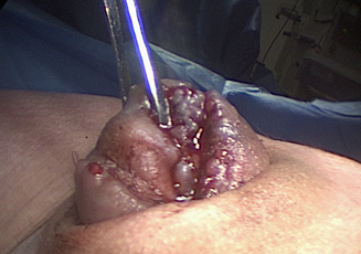
A 35-year-old patient presented to the gynaecology clinic with a history of worsening pelvic pain and cyclical umbilical bleeding on a background of grade 4 endometriosis, having undergoing a laparoscopy and excision of the endometriosis 2 years previously. Examination revealed a 3cm tender nodule in the umbilicus, a fixed uterus and retro-cervical tenderness. Subsequent MRI suggested an umbilical endometriotic nodule with retro-cervical endometriosis and findings at the time of her laparoscopy were of grade 4 endometriosis and a 3cm umbilical endometriotic nodue (Figure 1). Following laparoscopic excision of her pelvic endometriosis the umbilical nodule was excised. A circumferential incision was made around the nodule using diathermy, ensuring full excision with good margins. The underlying fascia was closed using 2/0 polyglactin (Vicryl ®) sutures and the umbilicus was then reconstructed using 2/0 poliglecaprone (Monocryl ®) sutures giving a good aesthetic result. The whole procedure was performed with laparoscopic assistance to ensure adequate excision and complete closure of the excised area. Histology confirmed endometriosis and follow up 6 months later confirmed a good aesthetic result with no evidence of recurrence.

Figure 1: Endometriotic umbilical nodue.
3. Discussion
Primary umbilical endometriosis, the more common form of umbilical endometriosis, develops spontaneously in the absence of any previous abdominal surgery. The exact pathogenesis is unknown, however possible theories include the spread of endometrial cells to the umbilicus through the abdominal cavity, via the lymphatic system or through the embryonic remnants in the umbilical folds [3-5]. Secondary umbilical endometriosis develops following previous surgery (caesarean section, abdominal hysterectomy, appendicectomy, laparoscopy etc.) Scholefield et al. [6] and is due to iatrogenic seeding of endometrial tissue. The clinical features include an umbilical swelling (90%), often associated with cyclical pain (81.5%) and bleeding (49.2%). The differential diagnoses include granuloma, umbilical polyps, haemangioma, melanocytic nevus, seborrhoeic keratosis, granular cell tumour and umbilical hernia. Investigations may include an ultrasound to assess the echogenicity and vascular involvement. MRI can also be helpful, although no imaging modality is truly diagnostic. Medical treatments such as the contraceptive pill or GnRH analogues are an option and can be effective in diminishing symptoms temporarily, however after cessation of hormonal treatment symptom recurrence is common [1]. The definitive management involves surgery, which can vary from superficial diathermy to the more radical omphalectomy with a concomitant laparoscopic approach. This allows for total excision of the umbilicus and repair of the underlying fascia and reconstruction of the umbilicus. Another option is local resection of the endometriotic tissue and sparing of the umbilicus. Whilst there is no real data on follow up of these surgically treated cases, Fedele et al. [7] described in their case series recurrence of umbilical endometriosis in those managed with superficial surgical approach [7]. They described a radical en bloc excision of the umbilicus including the fascia and perotineum, with subsequent histology confirming intrafascial endometriosis in all of those cases. This would support a more radical approach to surgery aided by laparoscopy to enable complete excision of umbilical endometriosis and reduce local recurrence. The use of laparoscopy also allows the extent of the nodule to be explored and ensures adequate excision of the lesion and the accurate reconstruction of the fascial planes, whilst also allowing concomitant pelvic disease to be treated.
Declaration of Interest
The authors report no declaration of interest
References
- Giudice LC, Kao LC. Endometriosis. Lancet 364 (2004): 1789-1799.
- Victory R, Diamond MP, Johns DA. Villar's nodule: a case report and systematic literature review of endometriosis externa of the umbilicus. J Minim Invasive Gynecol 14 (2007): 23-32.
- Efremidou EI, Kouklakis G, Mitrakas A, et al. Primary umbilical endometrioma: a rare case of spontaneous abdominal wall endometriosis. Int J Gen Med 5 (2012): 999-1002.
- Theunissen CIJM, Ijpma FFA. Primary umbilical endometriosis: a cause of a painful umbilical nodule. Journal of Surgical Case Reports (2015): rjv025.
- Fancellu A, Pinna A, Manca A, et al. Primary umbilical endometriosis. Case report and discussion on management options. Int J Surg Case Rep 4 (2013): 1145-1148.
- Scholefield HJ, Sajjad Y, Morgan PR. Cutaneous endometriosis and its association with caesarean section and gynaecological procedures. J Obstet Gynaecol 22 (2002): 553-554.
- Fedele L, Frontino G, Bianchi S, et al. Umbilical endometriosis: a radical excision with laparoscopic assistance. Int J Surg 8 (2010): 109-111.
