The effect of delaying composite resin restoration after silver diamine fluoride pretreatment on the shear bond strength of the final restoration: An in-vitro study
Article Information
Eman H. Aboulsaood1* BDS, Niveen S. Bakry1 PhD, Sarah I. Zeitoun1 PhD, Salma A. Aboulgheit2 PhD
1Pediatric Dentistry and Dental Public Health Department, Faculty of Dentistry, Alexandria University, Alexandria 21527, Egypt.
2Dental Biomaterials, Faculty of Dentistry, Faculty of Dentistry, Alexandria University, Alexandria 21527, Egypt.
Corresponding author: Eman Hossam el-din Aboulsaood, Resident at Pediatric Dentistry and Dental Public Health Department, Faculty of Dentistry, Alexandria University, Alexandria 21527, Egypt.
Received: January 31, 2023; Accepted: February 08, 2023;Published: February 23, 2023
Citation: Eman H. Aboulsaood BDS, Niveen S. Bakry PhD, Sarah I. Zeitoun PhD, Salma A. Aboulgheit PhD. The effect of delaying composite resin restoration after silver diamine fluoride pretreatment on the shear bond strength of the final restoration : An in-vitro study. Journal of Radiology and Clinical Imaging. 6 (2023): 32-40.
Share at FacebookAbstract
Background: The aim of the current study was to investigate the effect of delaying final restoration for one week after SDF pretreatment on shear bond strength of composite resin.
Materials and methods: Sixty-six extracted sound primary teeth were randomly allocated to one of three groups as follows ; Group 1 (Control group): composite restoration without pretreatment, group 2: 38% SDF pretreatment + immediate composite restoration, group 3: 38% SDF pretreatment + 1-week aging + composite restoration. Each group was further divided into two subgroups according to the bonding protocol used as follows: (Subgroup ER) etch-and-rinse technique and (Subgroup SE) self-etch technique. SDF and composite resin were applied to the flattened buccal dentinal surfaces. SBS was measured using universal testing machine. Mode of failure was assessed by stereomicroscope. Elemental contents and depth of penetration were assessed using energy dispersive x-ray stereoscopy (EDS). Results: A statistically significant difference in mean SBS was found between group 2 (39.88 ± 27.35) and group 1 (85.17 ± 40.43), but no significant difference was found between group 3 (96.70 ± 30.28) and group 1 (85.17 ± 40.43). Moreover, there was a statistically significant difference in mean SBS between subgroup 2ER (52.23 ± 30.13) and 2SE (27.53 ± 18.11). EDS analysis revealed deeper penetration of silver ions over time.
Conclusions: Delaying composite resin restoration bonding to primary dentin for one week after SDF pretreatment eliminated any negative impact on SBS of the final restoration.
Keywords
Silver diamine fluoride; Shear bond strength; Universal
adhesive; Etch-and-rinse technique; Self-etch technique; Delayed bonding
Article Details
Background
While traditional caries management philosophy is based on complete eradication of the affected tissues, causing irreversible destruction of valuable tooth structure [1,2], the modern model is redirected toward radical elimination of the disease and sustainable protection [3].The new modalities are usually cost-effective and demand minimal clinical set-up and personnel [4].
Silver Diamine Fluoride (SDF) become increasingly popular as a suitable alternative in situations where conventional complex treatment can be unfeasible[5]. It is a simple, affordable, non-invasive, rapid technique that efficiently controls pain and infection in young children with minimal caustic effects [6,7].
The unique advantages of SDF are derived from the combined effects of its components [5]. With silver and fluoride ions having double synergistic effects on hard tissues and bacteria. While fluoride’s main impact is the enhancement of remineralization [2, 8], silver ions have a dominant antibacterial effect [9, 10], in addition to inhibiting collagen degradation and forming insoluble compounds and granular spherical grains that precipitate in the inter-tubular areas [11,12]. This in turn ,increases microhardness and decreases dentinal sensitivity by plugging dentinal tubules, [12,13]. The added ammonium hydroxide stabilizes the level of alkalinity [8], which is crucial for arresting the progression of caries [2].
Nevertheless, SDF possesses several limitations. It results in black staining of biological matter of dental substrates due to the formation of metallic silver [14, 15]. To counter this effect, alternatives have been suggested, including the addition of potassium iodide (KI) to SDF, so it can react with any silver ions residues [16]. Ammonium hexafluorosilicate was also suggested as a potential substitute [17]. However, both options were not as effective as SDF in reducing lesion progression [17, 18]. The inability of SDF to adequately meet all aesthetic and functional demands, highlights the need to combine caries arrest with the use of a definitive restorative material whenever lesions are cavitated . This will also provide adequate sealing against bacteria and improve the cleansability of the tooth [19-21].
Tooth-colored fillings, including resin-based composites, are widely used in pediatric dentistry [22]. It can reach full setting immediately[23]. Bulk-fill composite has been introduced with a high curing depth of up to 5 mm to decrease polymerization shrinkage, improve marginal integrity of the restoration and expedite restorative intervention, which makes it an ideal choice for pediatric dentistry [24-26].
Variable dental adhesives were introduced to provide a durable intimate bond to tooth substrates [27,28]. Contemporary adhesive systems are based on etch-and-rinse and self-etching protocols [29]. The more recently introduced universal adhesives can serve as a multi-mode system [29, 30]. Bonding to dentin can be challenging due to its hydrated organic nature[27]. Recent adhesives have attempted to counter this by incorporating specific acidic and hydrophilic monomer molecules that superficially demineralize and infiltrate the dentin to achieve hybridization, which aims for a more reliable resin interlocking within moist surfaces. They are more time efficient and produce less sensitivity compared to total-etch technique [26, 31, 32].
Despite the significant advantages provided by combining SDF with a composite restoration, different studies have reported deleterious effects of the former on the shear bond strength (SBS) of the latter [22,33,34]. Different attempts were advocated to minimize this [21,33].
However, to date no studies have examined the effects of delaying composite restoration after SDF application on SBS of the final restoration. Therefore, the current study aimed to investigate the impact of prolonging the time-period between SDF application and the final restoration on shear bond strength of universal adhesive applied using different strategies. The first null hypothesis being tested was that SDF application had no effect on the SBS of universal adhesives bonded to the dentin of primary teeth. The second null hypothesis was that there would be no differences in the SBS of universal adhesives bonded to pretreated or un-pretreated primary dentin using the etch-and-rinse (ER) or self-etch (SE) protocols.
Material and Methods
Equipment
Micro applicator brushes, Plastic filling instrument, Diamond Disc, Dappen dish, Light Curing Unit (Bluephase G2 / Ivoclar Vivadent), Transparent Teflon Cylinder 2mm×5mm, Stereomicroscope, Thermocycling Unit,
Table 1: Materials used in the study.
|
Materials |
Manufacturer |
Composition |
PH |
|
38% Silver Diamine Fluoride |
Advantage arrest Elevate oral care, USA. |
Silver, fluoride, ammonia, FD, and C Blue#1, Deionized water. |
10-12 |
|
Acid etch |
Eco-Etch gel (Ivoclar Vivadent Inc., Schaan, Liechtenstein). |
37% phosphoric acid, water, pigments, silicon dioxide. |
0.1-0.4 |
|
Universal adhesive |
Tetric N-Bond universal (Ivoclar Vivadent Inc., Schaan, Liechtenstein). |
10-MDP, Bis-GMA, 2-Hydroxylethyl methacrylate (HEMA), dimethacrylates, methacrylate carboxylic acid polymer (MCAP), ethanol, water, silicon dioxide, initiator and stabilizer. |
2.5 |
|
Composite resin |
Tetric N- Ceram Bulk fill (Ivoclar Vivadent Inc., Schaan, Liechtenstein). |
Bis-GMA, Bis-EMA, urethane dimethacrylate (UDMA), fillers, fluoride salts, additives, catalyst, stabilizers, and pigments. |
|
|
Artificial saliva |
Prepared at faculty settings. |
200 gm Methyl Paraben, 10 gm Sodium carboxymethyl cellulose, 0.625 gm Potassium chloride, 0.059 gm Magnesium chloride, 0.166gm Dipotassium phosphate, 0.326 gm to 7 using NaOH2Potassium dihydrogen phosphate. Increased to 1 liter by adding H2O, pH adjusted |
7 |
Scanning Electron Microscope (Jeol JSM-IT200 InTouchScope TM).
Specimen preparation
This study included 66 sound extracted human primary teeth that were obtained from the Pediatric Dentistry Department at Alexandria University after collection of informed consent signatures from the legal guardians of the patients. The teeth were cleaned immediately after extraction and examined for the presence of caries, defects, or cracks using a light microscope at 20X magnification. The various steps of the study were carried out in accordance with CRIS guidelines and the relevant regulations. Ethical approval was obtained from the Research Ethics Committee, Faculty of Dentistry, Alexandria University, Egypt (IRB:0245– IORG:0008839). The study samples were stored in deionized water (DI) containing 0.01% thymol for use within six months.
The root of each specimen was cut below the cementoenamel Junction using a low-speed diamond disc, and the coronal part was embedded in 14 × 20 acrylic blocks. The buccal surface was abraded using silicon carbide 0.1 grit abrasive to create a flat bonding surface. A computer- generated list of random numbers (www.randomizer.org) was used to assign the specimens in blocks of 3 to one of the 3 study groups (n = 22), as follows:
Group 1 (control group): Composite resin was bonded on
a flattened dentin surface without pretreatment.
Group 2: Teeth were pretreated with 38% SDF, then composite resin was bonded immediately after to dentin.
Group 3: Teeth were first pretreated with 38% SDF, left in artificial saliva for seven days, and exposed to thermodynamic cycling, following which composite resin was bonded to the dentin.
Each group was further divided into two subgroups based on the protocol used in bonding the composite resin.
Subgroup ER: Etch-and-rinse technique Subgroup SE: Self-etch technique
SDF application
Thirty-eight percent SDF (Advantage arrest , Elevate oral care, USA) was applied to the dry flat dentin surface of teeth of test groups (2 and 3) using a micro applicator brush. Two consequent layers were applied, with each being left on the surface for 1 minute, then gently dried using compressed air [35].
Adhesive application
In the ER subgroup: the flat dentin surface of teeth was acid etched using 37% Phosphoric acid for 15 seconds, rinsed with water, then gently dried with air. Afterwards,
Tetric N-Bond universal® (Ivoclar Vivadent Inc., Schaan, Liechtenstein) was rubbed on the dentinal surface using a micro brush for 20 seconds, gently thinned with air for 5 seconds, and cured for 15 seconds[36]. Finally, composite resin application was carried out.
In the SE subgroup, the Tetric N-Bond universal® was applied directly to the flat dentin surface using the same procedure as subgroup ER. The composite resin was applied thereafter
Composite application
To create a standardized bonding area, a 2 × 5 plastic cylindrical-shaped mold was placed on the flat dentin surface in all groups. The Universal bond was applied using a micro- brush applicator, in accordance with the protocol for each subgroup. This was followed by introduction of the Tetric N-Ceram Bulk fill® composite to each plastic mold using a plastic filling instrument and curing for a period of 40 seconds [37].
Aging process
Immediately after SDF application, the teeth in group 3 were stored individually in closed containers filled with artificial saliva (37°C) for seven days. Each tooth was covered with 5 mm of artificial saliva which was replaced every 24 hours. After seven days, the specimens were subjected to thermodynamic cycling at 5°C to 55°C and 30 second dwell time for 20 minutes to mimic exposure to oral cavity conditions for one week.
Composite bonding was carried out in all study samples, and these were then subjected to 1000 thermodynamic cycles to mimic exposure to the oral cavity for one year [38].
SBS test
Sixty-six samples were tested by means of a rod of the universal testing machine (5ST, Tinuis Olsen, England) , at a cross-head speed of 0.5 mm/min until the bond fractured and the shear force was recorded. The force was measured in Newtons (N) and converted megapascals (MPa) to calculate SBS values[39].
Failure mode assessment
Failure mode was assessed using a stereomicroscope at a magnification of 25X, and was classified into: adhesive failure (between the composite resin and the primary dentin), cohesive failure (within the composite resin) and mixed [40].
Energy dispersive X-ray spectroscopy (EDS) analysis
The elemental content and penetration depth of SDF into the dentinal tubules over a period of one week were examined. Six teeth (numbered from 1 to 6) were assessed immediately after pretreatment and again after the time interval. The flat dentinal surfaces were pretreated using 38% SDF, as described previously. The teeth were then sectioned buccolingually using a micro-saw (Iso Met 4000), and dehydrated via immersion in ascending grades (50%, 70%, and 90%) of ethyl alcohol that was freshly prepared by diluting absolute alcohol with distilled water. The surfaces were examined under a scanning electron microscope (SEM; Jeol JSM-IT200 InTouchScopeTM) with EDS at an acceleration voltage of 20.0 kV without coating. Surface area analysis of nitrogen (N), fluoride (F), and silver (Ag) ion levels were performed at different points on the outer layer of the pretreated dentin of each tooth. The penetration depths of the same elements were also assessed. However, as silver penetration was not consistent, the maximum depth at which the element was detected was measured at ten different points and the mean value was calculated for each tooth. The assessment was repeated after one week of aging, and the concentration and average penetration depth of each element was measured again in the same manner.
Statistical Anlaysis
All data analyses were carried out using IBM SPSS for Windows (Version 23.0), and the significance level was set at p-value < 0.05. Descriptive statistics, plots, and normality tests were used to confirm normal distribution for all variables. Mean values and standard deviations (SD) were calculated for all numerical variables, while frequencies and percentages were calculated for categorical variables. Parametric tests were used for analysis, with comparison of failure modes between the study groups and subgroups being done using a Chi-squared test with a Monte Carlo corrected p-value (whenever indicated). Two-way ANOVA was used to assess the effects of the time delay and bonding techniques on composite SBS, and comparisons between the ER and SE techniques (subgroups) within each group were done using an independent samples t-test. Comparison of the EDS results at baseline and after one week were done using a paired t-test, and adjusted means, standard errors (SE), and 95% confidence intervals (CI) were calculated.
Table 2: Descriptive data for the study groups and subgroups.
|
Adjusted mean (SE) |
95% CI |
F of two- way ANOVA P value |
||
|
Group |
1 |
85.17 (7.04) b |
71.09, 99.25 |
F= 18.19 P <0.001* |
|
2 |
39.88 (7.04) a |
25.80, 53.96 |
||
|
3 |
96.70 (7.04) b |
82.62, 110.78 |
||
|
Subgroup |
ER |
78.90 (5.75) |
67.40, 90.39 |
F= 1.50 P= 0.23 |
|
SE |
68.93 (5.75) |
57.44, 80.43 |
||
SBS: Shear bond strength, SE: Standard Error, CI: Confidence Interval
*Statistically significant at p-value <0.05
a, b: Different letters denote statistically significant difference between groups using Bonferroni adjusted significance levels.
Results
Significant differences in mean SBS were encountered between the three main groups (p-value <0.001). Table 2 shows the descriptive statistics of SBS.
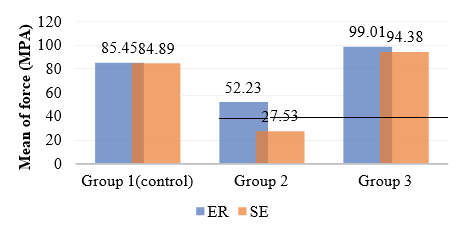
Upon comparison with group 1 (control group), a statistically significant difference in SBS was observed in group 2 (p-value < 0.001) ,but no significant difference was seen in group 3 (p-value = 0.76). Figure 1 shows the mean SBS values in the three groups and subgroups.
The results show that SBS of universal adhesive dropped significantly when composite resin was bonded immediately after SDF pretreatment, compared to the control group where samples were not pretreated. On the other hand, SDF had no negative impact on the SBS of universal adhesive when composite resin was bonded one week after pretreatment.
The independent t-test analysis showed a statistically significant difference in mean SBS between the ER (52.23 ± 30.13) and SE (27.53 ± 18.11) subgroups of group 2 only (p-value = 0.03). However, no significant differences in mean SBS were observed between the subgroups of the control group or group 3 (Table 3).
Failure mode
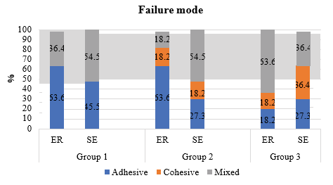
Figure 2 shows the failure modes observed in all samples in this study. The control group exhibited adhesive and mixed failures only. The ER subgroup predominantly exhibited adhesive failures in groups 1 and 2 and mixed failures in group 3. In contrast, the SE subgroup predominantly exhibited
Table 3: Mean SBS values by type of technique (etch-and-rinse and self-etch) and group
|
ER |
SE |
Difference |
Independent samples t-test p-value |
||
|
Mean ± SD |
(95% CI) |
||||
|
SBS (Mpa) |
Group 1 |
85.45 ± 33.75 |
84.89 ± 47.88 |
0.56 (-36.28, 37.41) |
0.98 |
|
Group 2 |
52.23 ± 30.13 |
27.53± 18.11 |
24.70 (2.59, 46.81) |
0.03* |
|
|
Group 3 |
99.01 ± 30.99 |
94.38 ± 30.87 |
4.63 (-22.88, 32.14) |
0.73 |
|
|
Mean |
78.90 ± 36.58 |
68.93 ± 44.90 |
10.08 (-10.17, 30.11) |
0.33 |
|
SBS: Shear bond strength, SD: Standard Deviation
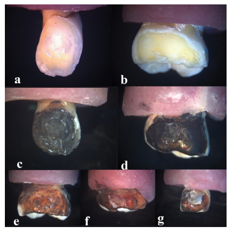
Figure 3: Failure modes in the 6 subgroups. (a) failure in control group was mainly adhesive in subgroup ER. (b) failure in control group was mainly mixed in subgroup SE. (c) failure in group 2 was mainly adhesive in subgroup ER. (d) Failure in group 2 was mainly mixed in subgroup SE. (e) Adhesive failure in group 3. (f) Mixed failure in group 3 (4) Cohesive failure in group 3.
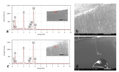
Figure 4: Illustration of SEM and EDS analysis of elemental contents for teeth pretreated with SDF in 2 occasions. (a) Atomic concentrations of the Ag,F,N on the most outer layer immediately pretreatment. (b) detection of silver element immediately after pretreatment. (c) reduction of atomic concentrations of the Ag,F,N on the most outer layer after 1 week of pretreatment. (d) detection of silver element at deeper levels after week.
mixed failures in groups 1 and 2, while all three types of failure were observed in group 3 without predominance (Figure 3). No significant differences in the type of failure at the composite resin level was observed between the study groups (p-value > 0.05).
EDS observations
Figure 4 shows the findings of the EDS analysis exploring the dentin concentrations of silver, fluoride, and nitrogen immediately after pretreatment and after aging. The average nitrogen ion atomic ratio was 3.42% immediately after pretreatment, and this decreased to 0.45% upon second examination. This difference was statistically significant (p-value = 0.002; Table 4).
The EDS and SEM observations showed deeper penetration of silver into dentinal tubules of sound dentin after one week of pretreatment (Figure 4). The mean maximum depth of penetration was 249.88 on the first analysis and increased to 871.45 after one week of pretreatment. Upon comparing depths on the two occasions, the difference was statistically significant (p-value=0.001;Table 5 ).
Discussion
This study investigated the SBS of composite resin bonded using two adhesion protocols to primary dentin that had been pretreated with 38% SDF. The findings showed that delaying composite resin application for one week after pretreatment significantly improved SBS when compared to application immediately after pretreatment. Significant differences in SBS were also observed between the ER and SE subgroups when composite was applied immediately after pretreatment; however, this difference disappeared upon delaying application. Based on these findings, both null hypotheses were partially rejected.
The current study implemented a delay of one week between restoration and SDF pretreatment. Ng et al., [21] previously hypothesized that separation of pretreatment and restoration would allow for SDF solidification and, consequently, enhance the SBS of the final restoration. However, the findings of their study showed no statistically significant differences between groups where glass ionomer restorations were placed immediately , and one week after SDF pretreatment [21].
It has to be noted that the dentin bonding mechanisms of glass ionomer and composite resin differ considerably, with the former using chemical adhesion dependent on weak polyacrylic acid and the latter using micromechanical adhesion [23, 41]. A systematic review by Frolich et al. (2022) concluded that although application of SDF did not affect the SBS of glass ionomer, it caused deterioration of the bonding systems [22]. In light of this data, composite resin was considered to be the material of choice as a final restoration after SDF pretreatment in the present study.
The findings of the present study showed that the SBS of composite resin significantly deteriorated when it was applied immediately after SDF application compared to the control group (p-value<0.001). This is consistent with the results obtained by Markham et al. [42] , Koizumi et al. [34] who used KI/SDF as a pretreatment, Kucukyilmaz et al. [43] who used SDF and ammonium hexafluorosilicate on intact and demineralized dentin and Lutgen et al., who pretreated sound dentin [33].
In contrast, Favaro et al. [44] and Quock et al. [19] reported no adverse effect of SDF on the composite micro-SBS and micro- tensile bond strength, respectively. Pérez-Hernández et al. [45] reported improvement in fissure sealant retention when applied after SDF application. This contradiction can be attributed to the fundamental differences between enamel and dentin nature, leading to the distinct impact of SDF and etching on each of the substrates [46-48].
SDF reaction with hydroxyapatite forms silver phosphate layer (Ag3PO4) [46]. The pretreatment also results in obstruction of dentinal tubules by silver particles [49]. The presence of this impermeable layer and inter-tubular precipitate acts as a phase separation interfering with intimate resin impregnation, which yields a suboptimal hybrid layer [22, 27, 33, 50]. Therefore, SBS of applied composite resin will be affected [22, 28]. Moreover, putting into consideration that SDF is a highly alkaline fluid (PH=10-12.5) [51]. It works as a physical and chemical barrier to adhesion that necessitates a dry acidic media to create a route for adhesive resin [33].
The significant improvement in SBS observed upon delaying composite restoration bonding to one week after pretreatment can be attributed to a deeper penetration of silver ions into the dentinal tubules during the time interval [21, 43, 46, 52]. In order to test this hypothesis, the maximum depth of penetration of silver was assessed immediately and one week after pretreatment using EDS. The findings showed that while silver could be detected at a depth of up to 309.5 µm initially, this increased to be up to 1001.1 µm when examined again one week after pretreatment. Moreover, this time interval also seemed to cause ammonia preservatives to leach out as nitrogen content significantly decreased.
Silver ions have an inhibitory effect on matrix metalloproteinases (MMPs) and cysteine cathepsin enzymes that degrade dentinal collagen matrix proteins, leading to bonding loss [53-55]. The re-mineralizing and inhibitory properties of SDF suggest it may have an enhancing effect on the SBS of composite resin if time interval were further extended.
With regard to the adhesion protocols used, a significant difference in mean SBS was observed between the two subgroups in group 2 only (p-value = 0.03).The obtained result is in line with Lutgen et al. [33] .This could potentially be attributed to the fact that the SE technique relies solely on functioning acidic monomers in universal adhesives, which exhibit mild acidity (pH = 2.5) that is considerably weaker compared to the 37% phosphoric acid (pH = 0.1–0.4) used in the ER [56, 57]. Those weak monomers are more likely to be buffered by the alkaline ammonia content of the SDF. Hence, more debonding events were encountered [33, 58].
Performance of SE subgroup was enhanced in group 3 when bonding was delayed to be similar to the ER subgroup, which provides an additional evidence that alkalinity of SDF decreases when left to set.
Assessment of failure mode was done using a stereomicroscope. Currently, there is a lack of consensus in the literature with regard to the classification of failure modes and the methodology of interpretation. The current study opted to use the Scherrers et al. classification [40], and found that the three groups did not significantly differ with regard to the type of failures observed between the composite restoration and the dentin.
The current study was limited by the inability to apply a conductive coating during SEM examination as the teeth were examined on two separate occasions. It should also be noted that only intact dentin was used in the current study, and the SBS is expected to be lower and the silver ion atomic ratio is anticipated to be higher in carious dentin [59, 60]. Further clinical studies are necessary to confirm these findings.
Conclusions
Within the limitations of this in-vitro study, it can be concluded that the application of SDF to primary dentin immediately before composite resin restoration adversely affects SBS regardless of the bonding protocol used. Therefore, SDF pretreatment one week prior to composite resin restoration is recommended to minimize the risk of bonding failure. The SE technique can be used in this situation without any deterioration of SBS.
List of Abbrevations
SDF: Silver diamine fluoride.
SBS: Shear bond strength.
ER: Etch-and-rinse
SE: Self-etch
10-MDP: 10- methacrylate carboxylic acid polymer Bis-GMA: Bisphenol A-glycidyl methacrylate
Bis-EMA: Bisphenol A-diglycidyl methacrylate ethoxylated HEMA: 2-Hydroxylethyl methacrylate
MCAP: Methacrylate carboxylic acid polymer UDMA: Urethane dimethacrylate
SD: Standard deviation. ANOVA: Analysis of variance.
EDS: Energy dispersive X-ray spectroscopy. MPa: Megapascals.
Declarations
Ethics approval and consent to participate
This study was completed in compliance with the Declaration of Helsinki. Ethical approval was obtained from the Research Ethics Committee, Faculty of Dentistry, Alexandria University, Egypt (IRB:0245–IORG 0008839). Informed consent was collected from the legal guardians of the patients prior to tooth extraction. All methods were performed in accordance with relevant guidelines and regulations.
Consent for publication
Not applicable
Availability of data and materials
The datasets used and/or analyzed in the current study are available from the corresponding author on reasonable upon request.
Competing interest
The authors declare that they have no competing interests
Funding
The authors did not receive any funds to support this research.
Authors' contributions
NSB developed the study concept; EHA executed the practical steps of the study,, interpreted the findings, and prepared the manuscript; SIZ and SA revised the practical steps of the study. All authors revised and approved the final manuscript prior to submission for publication.
Acknowledgements
Not applicable .
References
- Schwendicke F, Dörfer CE, Paris Incomplete caries removal: a systematic review and meta-analysis. J Dent Res. 92 (2013): 306-314.
- Mei ML, Lo ECM, Chu CH. Arresting Dentine Caries with Silver Diamine Fluoride: What's Behind It? J Dent 97 (2018): 751-758.
- Yon MJY, Gao SS, Chen KJ, Duangthip D, Lo ECM, et Medical Model in Caries Management. Dent J (Basel). 2019:7.
- Contreras V, Toro MJ, Elías-Boneta AR, Encarnación- Burgos Effectiveness of silver diamine fluoride in caries prevention and arrest: a systematic literature review. Gen Dent. 65 (2017): 22-29.
- Crystal YO, Niederman Evidence-Based Dentistry Update on Silver Diamine Fluoride. Dent Clin North Am. 63 (2019): 45-68.
- Sharma G, Puranik MP, K RS. Approaches to Arresting Dental Caries: An Update. J Clin Diagn Res. 9 (2015): Ze08-11.
- Seifo N, Cassie H, Radford JR, Innes Silver diamine fluoride for managing carious lesions: an umbrella review. BMC Oral Health. 19 (2019): 145.
- Zhao IS, Gao SS, Hiraishi N, Burrow MF, et Mechanisms of silver diamine fluoride on arresting caries: a literature review. Int Dent J. 68 (2018): 67-76.
- Policy on the Use of Silver Diamine Fluoride for Pediatric Dental Pediatr Dent. 39 (2017): 51-53.
- Horst JA, Ellenikiotis H, Milgrom PM. UCSF Protocol for Caries Arrest Using Silver Diamine Fluoride: Rationale, Indications and Consent. Pa Dent J (Harrisb). 84 (2017):16-26.
- Seto J, Horst JA, Parkinson DY, Frachella JC, DeRisi Enhanced Tooth Structure Via Silver Microwires Following Treatment with 38 Percent Silver Diamine Fluoride. Pediatr Dent. 42 (2020): 226-231.
- Mei ML, Ito L, Cao Y, Li QL, Lo EC, et Inhibitory effect of silver diamine fluoride on dentine demineralisation and collagen degradation. J Dent. 41 (2013): 809-817.
- Greenwall-Cohen J, Greenwall L, Barry Silver diamine fluoride - an overview of the literature and current clinical techniques. Br Dent J. 228 (2020): 831-838.
- Lou YL, Botelho MG, Darvell BW. Reaction of silver diamine fluoride with hydroxyapatite and J Dent. 39 (2011): 612-618.
- Yi Z, Ye J, Kikugawa N, Kako T, Ouyang S, et al. An orthophosphate semiconductor with photooxidation properties under visible-light irradiation. Nat Mater. 9 (2010): 559-564.
- Zhao IS, Chu S, Yu OY, Mei ML, Chu CH, et Effect of silver diamine fluoride and potassium iodide on shear bond strength of glass ionomer cements to caries-affected dentine. Int Dent J. 69 (2019): 341-347.
- Peng JJ, Botelho MG, Matinlinna JP. Silver compounds used in dentistry for caries management: a J Dent. 40 (2012): 531-541.
- Turton B, Horn R, Durward C. Caries arrest and lesion appearance using two different silver fluoride therapies on primary teeth with and without potassium iodide: 12-month Clin Exp Dent Res. 7 (2021): 609-619.
- Quock RL, Barros JA, Yang SW, Patel SA. Effect of silver diamine fluoride on microtensile bond strength to Oper Dent. 37 (2012): 610-616.
- Mei ML, Zhao IS, Ito L, Lo EC, Chu CH. Prevention of secondary caries by silver diamine Int Dent J. 66 (2016): 71-77.
- Ng E, Saini S, Schulze KA, Horst J, Le T, et Shear Bond Strength of Glass Ionomer Cement to Silver Diamine Fluoride-Treated Artificial Dentinal Caries. Pediatr Dent. 42 (2020): 221-225.
- Fröhlich TT, Botton G, Rocha RO. Bonding of Glass- Ionomer Cement and Adhesives to Silver Diamine Fluoride-treated Dentin: An Updated Systematic Review and Meta-Analysis. The journal of adhesive dentistry. 24 (2022): 29-38.
- Sauro S, Faus-Matoses V, Makeeva I, Nuñez Martí JM, Gonzalez Martínez R, et al. Effects of polyacrylic acid pre-treatment on bonded-dentine interfaces created with a modern bioactive resin-modified glass ionomer cement and subjected to cycling mechanical Materials (Basel). 11 (2018):1884.
- Paganini A, Attin T, Tauböck TT. Margin Integrity of Bulk-Fill Composite Restorations in Primary Materials (Basel). 2020; 13.
- Akman H, Tosun Clinical evaluation of bulk-fill resins and glass ionomer restorative materials: A 1-year follow- up randomized clinical trial in children. Niger J Clin Pract. 23 (2020): 489-497.
- Rosa WL, Piva E, Silva AF. Bond strength of universal adhesives: A systematic review and meta-analysis. J 43 (2015): 765-776.
- Tjäderhane Dentin bonding: can we make it last? Oper Dent. 40 (2015): 4-18.
- Marshall GW, Jr., Marshall SJ, Kinney JH, Balooch M. The dentin substrate: structure and properties related to J Dent. 25 (1997): 441-458.
- Sofan E, Sofan A, Palaia G, Tenore G, Romeo U, Migliau
- Classification review of dental adhesive systems: from the IV generation to the universal type. Ann Stomatol (Roma). 8 (2017): 1-17.
- Cardoso GC, Nakanishi L, Isolan CP, Jardim PDS, Moraes Bond Stability of Universal Adhesives Applied To Dentin Using Etch-And-Rinse or Self-Etch Strategies. Braz Dent J. 30 (2019): 467-475.
- Proença JP, Polido M, Osorio E, Erhardt MC, Aguilera FS, et al. Dentin regional bond strength of self-etch and total-etch adhesive systems. Dental materials : official publication of the Academy of Dental 23 (2007): 1542-1548.
- Manuja N, Nagpal R, Pandit Dental adhesion: mechanism, techniques and durability. J Clin Pediatr Dent. 36 (2012): 223-234.
- Lutgen P, Chan D, Sadr A. Effects of silver diammine fluoride on bond strength of adhesives to sound dentin. Dent Mater 37 (2018): 1003-1009.
- Koizumi H, Hamama HH, Burrow MF. Effect of a silver diamine fluoride and potassium iodide-based desensitizing and cavity cleaning agent on bond strength to dentine. International Journal of Adhesion and 68 (2016):54-61.
- Hu S, Meyer B, Duggal A silver renaissance in dentistry. Eur Arch Paediatr Dent. 19 (2018): 221-227.
- Loguercio AD, Luque-Martinez IV, Fuentes S, Reis A, Muñoz MA. Effect of dentin roughness on the adhesive performance in non-carious cervical lesions: A double- blind randomized clinical J Dent. 69 (2018): 60-69.
- Han SH, Park SH. Incremental and Bulk-fill Techniques With Bulk-fill Resin Composite in Different Cavity Oper Dent. 43 (2018): 631-641.
- Zanatta RF, Lungova M, Borges AB, Torres C, Sydow HG, et al. Microleakage and Shear Bond Strength of Composite Restorations Under Cycling Conditions. Oper 42 (2017): E71-e80.
- Klaisiri A, Krajangta N, Thamrongananskul The Durability of Zirconia/Resin Composite Shear Bond Strength using Different Functional Monomer of Universal Adhesives. Eur J Dent. 2021.
- Scherrer SS, Cesar PF, Swain Direct comparison of the bond strength results of the different test methods: a critical literature review. Dental materials : official publication of the Academy of Dental Materials. 26 (2010): e78-93.
- Khan AR, Fida M, Gul Decalcification and bond failure rate in resin modified glass ionomer cement versus conventional composite for orthodontic bonding: A systematic review & meta-analysis. Int Orthod. 18 (2020): 32-40.
- Markham MD, Tsujimoto A, Barkmeier WW, Jurado CA, Fischer NG, et al. Influence of 38% silver diamine fluoride application on bond stability to enamel and dentin using universal adhesives in self-etch mode. Eur J Oral Sci.128 (2020): 354-360.
- Kucukyilmaz E, Savas S, Akcay M, Bolukbasi B. Effect of silver diamine fluoride and ammonium hexafluorosilicate applications with and without Er:YAG laser irradiation on the microtensile bond strength in sound and caries- affected Lasers Surg Med. 48 (2016): 62-69.
- Favaro JC, de Mello Peixoto YCT, Geha O, Dias FA, Guiraldo RD, et al. Can silver diamine fluoride or silver nanoparticle-based anticaries agents to affect enamel bond strength? Restor Dent 46 (2021): e7.
- Pérez-Hernández J, Aguilar-Díaz FC, Venegas-Lancón RD, Gayosso CAÁ, Villanueva-Vilchis MC, et al. Effect of silver diamine fluoride on adhesion and microleakage of a pit and fissure sealant to tooth enamel: in vitro trial. Eur Arch Paediatr 19 (2018): 411-416.
- Li Y, Liu Y, Psoter WJ, Nguyen OM, Bromage TG, et . Assessment of the Silver Penetration and Distribution in Carious Lesions of Deciduous Teeth Treated with Silver Diamine Fluoride. Caries Res. 53 (2019): 431-440.
- Giannini M, Makishi P, Ayres AP, Vermelho PM, Fronza BM, et Self-etch adhesive systems: a literature review. Braz Dent J. 26 (2015): 3-10.
- Jacker-Guhr S, Sander J, Luehrs AK. How "Universal" is Adhesion? Shear Bond Strength of Multi-mode Adhesives to Enamel and Dentin. The journal of adhesive dentistry. 21 (2019): 87-95.
- Sulyanto RM, Kang M, Srirangapatanam S, Berger M, Candamo F, et al. Biomineralization of Dental Tissues Treated with Silver Diamine Fluoride. J Dent Res. 2021; 100 (2021): 1099-1108.
- Pashley DH, Tay FR, Carvalho RM, Rueggeberg FA, Agee KA, et From dry bonding to water-wet bonding to ethanol-wet bonding. A review of the interactions between dentin matrix and solvated resins using a macromodel of the hybrid layer. Am J Dent. 20 (2007): 7-20.
- Sayed M, Matsui N, Hiraishi N, Inoue G, Nikaido T, et. Evaluation of discoloration of sound/demineralized root dentin with silver diamine fluoride: In-vitro study. Dent Mater J. 38 (2019): 143-149.
- Seto J, Horst JA, Parkinson DY, Frachella JC, DeRisi JL. Silver microwires from treating tooth decay with silver diamine BioRxiv. 2017:152199.
- Kiuru O, Sinervo J, Vähänikkilä H, Anttonen V, Tjäderhane MMP Inhibitors and Dentin Bonding: Systematic Review and Meta-Analysis. Int J Dent. 2021:9949699.
- Mei ML, Ito L, Cao Y, Li QL, Chu CH, et The inhibitory effects of silver diamine fluorides on cysteine cathepsins. J Dent. 2014; 42 (2014): 329-335.
- Mei ML, Ito L, Cao Y, Lo ECM, Li QL, et An ex vivo study of arrested primary teeth caries with silver diamine fluoride therapy. J Dent. 42 (2014): 395-402.
- Escobar M, Souza JCM, Barra GMO, Fredel MC, Özcan M, et al. On the synergistic effect of sulfonic functionalization and acidic adhesive conditioning to enhance the adhesion of PEEK to resin-matrix composites. Dental materials : official publication of the Academy of Dental Materials. 37 (2021): 741-754.
- Pashley DH, Tay FR, Breschi L, Tjäderhane L, Carvalho RM, et State of the art etch-and-rinse adhesives. Dental materials : official publication of the Academy of Dental Materials. 27 (2011): 1-16.
- Carvalho RM, Chersoni S, Frankenberger R, Pashley DH, Prati C, et al. A challenge to the conventional wisdom that simultaneous etching and resin infiltration always occurs in self-etch adhesives. Biomaterials. 26 (2005): 1035-1042.
- Kaup M, Dammann CH, Schäfer E, Dammaschke Shear bond strength of Biodentine, ProRoot MTA, glass ionomer cement and composite resin on human dentine ex vivo. Head Face Med. 2015; 11:14.
- Sayed M, Matsui N, Uo M, Nikaido T, Oikawa M, et Morphological and elemental analysis of silver penetration into sound/demineralized dentin after SDF application. Dental materials : official publication of the Academy of Dental Materials. 35 (2019): 1718-1727.


