STEMI Coexisted with COVID 19 in a 35-Year-Old Patient
Article Information
Kawtar Manouri1*, Rida Touab2, Nour Elhouda Snoussi1, Yousra Oussou1, Nawal Doghmi1
1Department of Cardiology B, IBN Sina Hospital Center, Rabat, Morocco
2Department of Anesthesiology and critical care, IBN Sina Hospital Center, Rabat, Morocco
*Corresponding author: Kawtar Manouri, Department of Cardiology B, IBN Sina Hospital Center, Rabat, Morocco
Received: 23 March 2021; Accepted: 31 March 2021; Published: 30 June 2021
Citation: Kawtar Manouri, Rida Touab, Nour Elhouda Snoussi, Yousra Oussou, Nawal Doghmi. STEMI Coexisted with COVID 19 in a 35-Year-Old Patient. Cardiology and Cardiovascular Medicine 5 (2021): 391-394.
Share at FacebookAbstract
Background: Corona viruses are known to attack the cardiovascular system, and it also appears that the virus may attack the heart muscle directly.
Case presentation: We report the case of a 35 year-old-woman admitted for respiratory distress related to COVID 19 infection complicated to myocardial infarction with non-obstructive coronary arteries.
Conclusion: The distinction between ischemic and non-ischemic causes of cardiac injury remains difficult given the limitation of additional examinations in this particular context, which maintains confusion in the identification of these breaches.
Keywords
Covid-19; STEMI; Myocardial injury; MINOCA
Article Details
1. Introduction
The 2019 coronavirus pandemic (COVID-19) affected all aspects of cardiology practice. The contribution of each of the suspected causes to myocardial lesions in this context remains difficult to determine. The initial presentation and electrocardiogram (ECG) changes in this situation can resemble with ST elevation myocardial infarction (STEMI).
2. Case Report
We report the case of a 35-year-old woman without cardiovascular risk factors or specific medical history, admitted to emergency room for respiratory distress. Two days prior to admission, she had acute constrictive chest pain radiating to the left shoulder. It was complicated by respiratory discomfort following the use of no steroidal anti-inflammatory drugs. Physical examination on admission revealed a polypneal patient with signs of respiratory distress. Oxygen saturation as measured by pulse oximetry [SpO2] was 50%. The patient was tachycardic; her rate was 140 beats per minute (bpm).
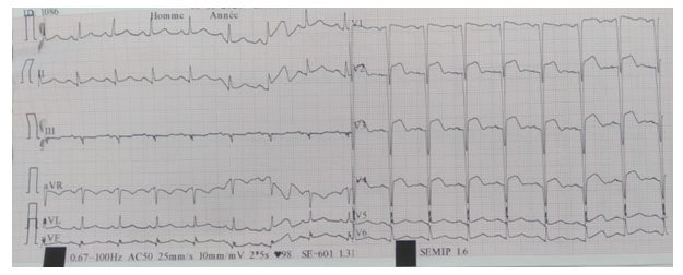
The electrocardiogram (ECG) recorded sinus tachycardia with anterior wall STEMI (Figure 1). A thoracic CT scan was in favour of COVID 19 infection with 75% lung parenchyma involvement. Laboratory investigations at the time of diagnosis yielded the following results: HGB: 13.4 g/dl, WBC: 17910 / mm³, PLT: 549000 /mm³, Urea: 0.3 g/l, Creatinine: 7 mg/l, C-Reactive Protein: 352 mg/l, Na+: 141 mEq/l, K+: 4.2 mEq/l. The troponin was greater than 50.000 ng/l. The transthoracic ultrasound showed a left ventricle of borderline size with segmental kinetic disorders: severe hypokinesis of the septal, anterior and anterolateral walls with left ventricular dysfunction at 35%. Heart valves were without significant changes.
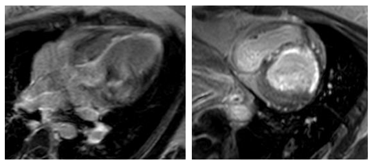
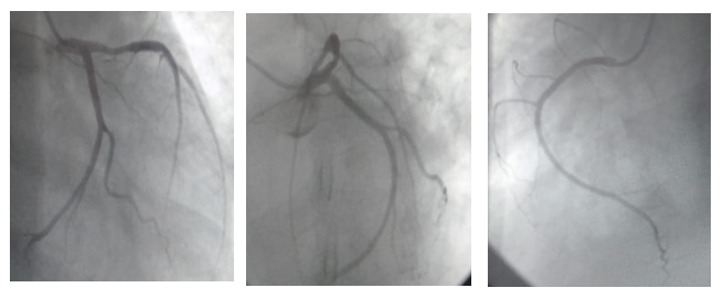
There was no fluid in the pericardial cavity. The patient was given non-invasive ventilation with treatment of ischemic heart disease, antibiotic therapy and hydroxychloroquine with good progression. Cardiac MRI carried out after stabilisation and discharge of the patient confirmed the sequelae of necrosis in the area of Left Anterior Descending (LAD) artery presumed to be partially viable (Figure 2). The coronary angiography was in favour of a balanced atheromatous coronary network, free of angiographically significant lesions (Figure 3).
3. Discussion
Coronavirus disease of 2019 (COVID-19) caused by infection from severe acute respiratory syndrome coronavirus 2 (SARS-CoV-2), is a cause of significant morbidity and mortality worldwide. The COVID-19 virus (SARS-CoV-2), uses the angiotensin converting enzyme (ACE) 2 for entry into target cells. ACE2 is predominantly expressed by epithelial cells of the lung, intestine, kidney, heart, and blood vessels. Acute cardiac injury associated or not with electric modifications is commonly described [1, 2]. In clinical practice, when myocardial injury is associated with typical chest pain, electrocardiographic and echocardiographic abnormalities, an acute coronary syndrome should be suspected. If coronary artery disease is eliminated alternative causes of myocardial infarction with non-obstructive coronary arteries (MINOCA) should be investigated. In non ischemic causes, the hypothesis that SARS-CoV-2 can be at the origin of acute myocarditis must be rapidly suspected.
In our patient cardiac MRI has objectified the sequelae of necrosis in the area of Left Anterior Descending (LAD) artery presumed to be partially viable but the coronary angiography was in favour of a balanced atheromatous coronary network, free of angiographically significant lesions. A deep hypo-xemia associated with a tachycardia may cause chest pain and electrocardiographic changes of myocardial ischemia. When the biomarkers are altered, myocardial infarction type 2 can be suggested. The mechanisms of acute cardiac injury in our patient may be direct myocardial injury by the virus through ACE2 entry, hypoxia-induced myocardial injury, microvascular damage and endothelial shedding, and cytokine/ inflammation-mediated damage [3]. These conditions can coexist in the same patients. Very varies and multisystemic clinical presentations observed in patients leave to think that the most serious forms of affection more like an inflammatory disease severe as a viral infection [4]. The SAR-SCov2 may cause an intense immune response with significant release of pro-inflammatory cytokines [2, 5, 6] which can lead to a ventricular dysfunction [7].
5. Conclusion
Elevation of cardiac biomarkers, particularly high-sensitivity troponin, is common in patients with COVID-19 infection, comprehensive cardiovascular examinations after balancing the risk of infection are crucial for differential diagnosis and precise treatment.
References
- Chaolin Huang, Yeming Wang, Xingwang
Li, Lili Ren, Jianping Zhao, Yi Hu, et al. Clinical features of patients infected with 2019 novel coronavirus in Wuhan, China. Lancet 395 (2020): 497-506.
- Fei Zhou, Ting Yu, Ronghui Du, Guohui Fan, Ying Liu, Zhibo Liu, et al. Clinical course and risk factors for mortality of adult inpatients with COVID-19 in Wuhan, China: a retrospective cohort study. Lancet 395 (2020): 1054-1062.
- Shi S, Qin M, Shen B, Cai Y, Liu T, Yang F, et al. Association of Cardiac Injury with mortality in hospitalized patients with COVID- 19 in Wuhan, China. JAMA Cardiol (2020): 1-8.
- Christophe Scavee, Agnes Pasquet, Christophe Beauloye. Aspects cardiologiques de l’infection par le COVID-19. Louvain Med 2020 mai-juin 139 (2020) : 236-246.
- Puja Mehta, Daniel F McAuley, Michael Brown, Emilie Sanchez, Rachel S Tattersall, Jessica J Manson, et al. COVID-19: consider cytokine storm syndromes and immunosuppression. Lancet Lond Engl 395 (2020): 1033-1034.
- Stanciu AE. Cytokines in heart failure. Adv Clin Chem 93 (2019): 63-113.
- Tao Guo, Yongzhen Fan, Ming Chen, Xiaoyan Wu, Lin Zhang, Tao He, et al. Cardiovascular implications of fatal outcomes of patients with Coronavirus Disease 2019 (COVID 19). JAMA Cardiol 5 (2020): 811-818.


