Solitary Fibrous Tumour of Pelvic Soft Tissue Mimicking A Prostatic Mass: Diagnosis and Management
Article Information
Keval N Patel* and Shashank J Pandya
Department of Surgical Oncology, The Gujarat Cancer Research Institute, Ahmedabad, Gujarat, India
*Corresponding Author: Keval N Patel, Department of Surgical Oncology, The Gujarat Cancer Research Institute, Ahmedabad, Gujarat, India, Tel: +919427052870;
Received: 10 October 2017; Accepted: 13 November 2017; Published: 16 November 2017
Share at FacebookAbstract
Solitary fibrous tumor (SFT) is a rare soft tissue tumor occurring rarely in extrapleural sites like the pelvis. We here present a case of SFT of the pelvis which was initially diagnosed radiologically as a mass arising from prostate and on biopsy found to be a solitary fibrous tumor. On exploration, there was a well encapsulated tumor in retro-vesical space separate from prostate. Histopathology with Immuno-histochemistry confirmed the diagnosis of SFT. We retrospectively reviewed the literature to avoid such confusion in future. Pubmed search was made with the following keywords: solitary fibrous tumor, treatment, soft tissue, pelvis, radiology, and histopathology.
Keywords
Solitary fibrous tumor, Treatment, Soft tissue, Pelvis, Radiology, Histopathology
Solitary fibrous tumor articles Solitary fibrous tumor Research articles Solitary fibrous tumor review articles Solitary fibrous tumor PubMed articles Solitary fibrous tumor PubMed Central articles Solitary fibrous tumor 2023 articles Solitary fibrous tumor 2024 articles Solitary fibrous tumor Scopus articles Solitary fibrous tumor impact factor journals Solitary fibrous tumor Scopus journals Solitary fibrous tumor PubMed journals Solitary fibrous tumor medical journals Solitary fibrous tumor free journals Solitary fibrous tumor best journals Solitary fibrous tumor top journals Solitary fibrous tumor free medical journals Solitary fibrous tumor famous journals Solitary fibrous tumor Google Scholar indexed journals Treatment articles Treatment Research articles Treatment review articles Treatment PubMed articles Treatment PubMed Central articles Treatment 2023 articles Treatment 2024 articles Treatment Scopus articles Treatment impact factor journals Treatment Scopus journals Treatment PubMed journals Treatment medical journals Treatment free journals Treatment best journals Treatment top journals Treatment free medical journals Treatment famous journals Treatment Google Scholar indexed journals Soft tissue articles Soft tissue Research articles Soft tissue review articles Soft tissue PubMed articles Soft tissue PubMed Central articles Soft tissue 2023 articles Soft tissue 2024 articles Soft tissue Scopus articles Soft tissue impact factor journals Soft tissue Scopus journals Soft tissue PubMed journals Soft tissue medical journals Soft tissue free journals Soft tissue best journals Soft tissue top journals Soft tissue free medical journals Soft tissue famous journals Soft tissue Google Scholar indexed journals Pelvis articles Pelvis Research articles Pelvis review articles Pelvis PubMed articles Pelvis PubMed Central articles Pelvis 2023 articles Pelvis 2024 articles Pelvis Scopus articles Pelvis impact factor journals Pelvis Scopus journals Pelvis PubMed journals Pelvis medical journals Pelvis free journals Pelvis best journals Pelvis top journals Pelvis free medical journals Pelvis famous journals Pelvis Google Scholar indexed journals Radiology articles Radiology Research articles Radiology review articles Radiology PubMed articles Radiology PubMed Central articles Radiology 2023 articles Radiology 2024 articles Radiology Scopus articles Radiology impact factor journals Radiology Scopus journals Radiology PubMed journals Radiology medical journals Radiology free journals Radiology best journals Radiology top journals Radiology free medical journals Radiology famous journals Radiology Google Scholar indexed journals Histopathology articles Histopathology Research articles Histopathology review articles Histopathology PubMed articles Histopathology PubMed Central articles Histopathology 2023 articles Histopathology 2024 articles Histopathology Scopus articles Histopathology impact factor journals Histopathology Scopus journals Histopathology PubMed journals Histopathology medical journals Histopathology free journals Histopathology best journals Histopathology top journals Histopathology free medical journals Histopathology famous journals Histopathology Google Scholar indexed journals prostatic articles prostatic Research articles prostatic review articles prostatic PubMed articles prostatic PubMed Central articles prostatic 2023 articles prostatic 2024 articles prostatic Scopus articles prostatic impact factor journals prostatic Scopus journals prostatic PubMed journals prostatic medical journals prostatic free journals prostatic best journals prostatic top journals prostatic free medical journals prostatic famous journals prostatic Google Scholar indexed journals cystoprostatectomy articles cystoprostatectomy Research articles cystoprostatectomy review articles cystoprostatectomy PubMed articles cystoprostatectomy PubMed Central articles cystoprostatectomy 2023 articles cystoprostatectomy 2024 articles cystoprostatectomy Scopus articles cystoprostatectomy impact factor journals cystoprostatectomy Scopus journals cystoprostatectomy PubMed journals cystoprostatectomy medical journals cystoprostatectomy free journals cystoprostatectomy best journals cystoprostatectomy top journals cystoprostatectomy free medical journals cystoprostatectomy famous journals cystoprostatectomy Google Scholar indexed journals Immuno-histochemistry articles Immuno-histochemistry Research articles Immuno-histochemistry review articles Immuno-histochemistry PubMed articles Immuno-histochemistry PubMed Central articles Immuno-histochemistry 2023 articles Immuno-histochemistry 2024 articles Immuno-histochemistry Scopus articles Immuno-histochemistry impact factor journals Immuno-histochemistry Scopus journals Immuno-histochemistry PubMed journals Immuno-histochemistry medical journals Immuno-histochemistry free journals Immuno-histochemistry best journals Immuno-histochemistry top journals Immuno-histochemistry free medical journals Immuno-histochemistry famous journals Immuno-histochemistry Google Scholar indexed journals articles Research articles review articles PubMed articles PubMed Central articles 2023 articles 2024 articles Scopus articles impact factor journals Scopus journals PubMed journals medical journals free journals best journals top journals free medical journals famous journals Google Scholar indexed journals
Article Details
1. Introduction
SFT is a rare soft tissue tumor and even rarer in pelvic soft tissue [1]. The knowledge of extraprostatic versus prostatic site of origin can avoid unnecessary cystoprostatectomy and its related morbidities especially when a tumor is large in size as it is very difficult to diagnose the site of origin based on available preoperative parameters. We retrospectively analyzed the factors which may lead to the diagnosis of the tumor site preoperatively and precautions to be taken during surgery if the site of origin is not known.
2. Case Description
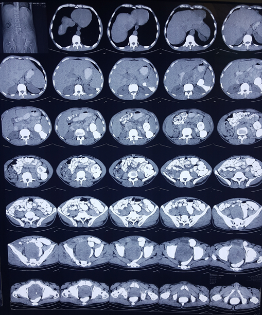
A 58 years old male patient presented with obstructive LUTS lasting for 3 months duration with increasing severity till he developed retention. On DRE there was grade III enlargement of prostate which was nodular, nontender with obliterated midline sulcus. His laboratory investigations were as follows: creatinine 1.0 mg/dl, PSA 0.8 ng/ml, urine culture showed no growth of organisms and LFT were within normal limits. CT scan of abdomen and pelvis showed
15 × 10 cm mass lesion arising from prostate without significant lymphadenopathy (Figure 1). TRUS guided biopsy showed mesenchymal tumour. Subsequently IHC was done which was suggestive of SFT (vimentin+, desmin+, CD34+). Institutional tumour board discussion suggested upfront surgery in view of localised disease. On exploration the tumour was approached first and dissected from all sides before approaching the bladder pedicles. The tumour was separate from prostate and bladder and was arising from soft tissue of pelvis. Post operative period was uneventful and patient was discharged on 4th post operative day. Histopathological examination revealed Solitary Fibrous Tumour confirmed on IHC with positivity for actin, vimentin, desmin, CD34, S100 and AE1. The patient is in followup for 1 year now and is recurrence free.

Figure 1: Shows large mass arising from pelvis, prostate not seen separately.
3. Discussion
SFT is a rare tumor, originally described in pleura [1]. It is rarer in retroperitoneum and less than 30 cases have been reported in the literature [2]. SFT of pelvic soft tissue usually occurs in retrovesical or perivesical space. It can be easily mistaken for a mesenchymal tumor arising from prostate especially if a tumour is large and occupies the whole pelvis making it difficult to diagnose the site of origin. We retrospectively tried to analyze the factors that could help us in diagnosing the site of origin for better planning and management.
Clinically pelvic SFT can be abnormally large in size producing symptoms due to compression of surrounding organs [3]. It is usually painless unless pain caused by compression of surrounding nerves. It may present as dysuria, acute urinary retention, hydroureteronephrosis constipation or even isolated pollakiuria [4, 5]. Bearing the fact that mass arising from the prostate may present with any of the above-mentioned symptoms, it is very difficult to diagnose the site based on clinical findings alone [6]. Serum PSA level is perceived to help in the diagnosis of the mass arising from the prostate may have high levels but the reliability is doubtful as in certain varieties of prostatic malignancy like the neuroendocrine differentiation, sarcoma or prostatic SFT it remains normal.
Radiologically CT scan in a case of pelvic SFT may show large mass with compression of surrounding organs. Wignall et al concluded that 56% of SFT had a size of >10 cm, all had discrete margins with majority had lobulated contours and heterogeneous strong enhancement. The local invasion was found in 9% of cases [7]. Theoretically, SFTs tend to be well encapsulated with preserved fat planes unless they are locally invasive. However definite criteria to differentiate it from a mass arising from prostate are not very well defined. Yiming et al have reported that prostatic SFT may present with a diameter of 4.7?25 cm average size being 9.8 cm. So even prostatic SFT can present as large mass.
Biopsy of a tumour provides definitive histological diagnosis although it may not differentiate between prostatic and extraprostatic site. Fletcher et al described the classical histopathological findings of SFT with positive results of immunohistochemical testing for Bcl-2 and vimentin and negative results for c-kit [8]. However, Pins et al in their study concluded that prostatic SFTs appear similar to those identified in extraprostatic sites and admixed prostatic tissue is not commonly associated with these lesions [9]. The high rate of recurrence has been reported in cases of prostatic SFT which were managed by TURP since it doesn’t involve complete removal of a tumour [10]. So biopsy is required in all cases of large mass arising in pelvis even if the radiological evidence shows it arising from prostate and PSA is normal.
The management of prostatic SFT includes radical prostatectomy and cystoprostatectomy if the mass is large and infiltrates bladder. Organ preserving complete surgical resection with pathologically negative surgical margins is the aim in case of SFT for good prognosis and since they are well encapsulated in a majority of the cases [1]. So on an exploration of any large pelvic SFT, the tumor should be approached first and dissected from all sides outside the capsule before dealing with the vesical pedicles to avoid inadvertent ligation and thus unnecessary
cystoprostatectomy. The bladder should be removed only if a tumour is locally invasive and it is not possible to develop planes between it and the tumor [11]. Some cases of SFT may have excess vascularity which may lead to torrential hemorrhage and so these should be handled with meticulous care [12]. Fahd-Aghale et al suggested pre operative embolization in large infiltrating SFT of a pelvis, however, could not make a clear statement because of a low incidence of such tumors [11].
4. Conclusion
SFT of the soft tissue of pelvis is a rare tumor. If a tumour is large, it is not possible to determine the site of origin (prostatic or extraprostatic) based on clinical features, biochemical investigations, radiological features or even biopsy. However preoperative biopsy is advisable in all cases of large masses even if the PSA is normal. On exploration, it is advisable to approach a tumour first and from all sides to avoid inadvertent ligation of vesical pedicles and thus cystoprostatectomy.
References
- Gold JS, Antonescu CR, Hajdu C, et al. Clinicopathologic correlates of solitary fibrous tumors. Cancer 94 (2002): 1057-1068.
- Saint-Blancard P, Jancovici R. Solitary fibrous tumor of the retroperitoneum. Rev Med Interne 30 (2009): 181-185.
- Hobarth K, Hofbauer J, Wrba F. Malignant hemangiopericytoma of the pelvis. Report of a case with urological implications and immunohistochemical analysis. Urol Int 47 (1991): 94-97.
- Rosenblatt SG. Walsh JC, Basile JJ. Pelvic hemangiopericytoma in a patient with mixed germ cell tumor of the testis. J Urol 166 (2001): 1824-1825.
- Tsushimi. Retroperitoneal solitary fibrous tumor of the pelvis with pollakiuria: a case report. BMC Research Notes 5 (2012): 593.
- Yiming Gu, Haijian Wei, Chunshu Rong, et al. Prostate solitary fibrous tumor: a case report and review of literature. Int J Clin Exp Pathol 9 (2016): 9675-9680.
- Wignall OJ, Moskovic EC, Thway K, et al. Solitary fibrous tumors of the soft tissues: review of the imaging and clinical features with histopathologic correlation. AJR Am J Roentgenol 195 (2010): W55-W62.
- Fletcher CDM, Unni K, Meryens F. World Health Organization classification of tumors, pathology and genetics, tumors of soft tissue and bone. Lyon: IARC Press (2002): 86-88.
- Pins MR, Campbell SC, Laskin WB, et al. Solitary fibrous tumor of the prostate: a report of 2 cases and review of the literature. Arch Pathol Lab Med 125 (2001): 274-277.
- Fard-Aghaie M, Stavrou GA. Honarpisheh H, et al. Large hemangiopericytoma of the pelvis?towards a multidisciplinary approach. World Journal of Surgical Oncology 13 (2015): 261.
- Andrea BG, Mazzucchelli R, Scarpelli M, et al. Solitary Fibrous Tumour of the Prostate Identified on Needle Biopsy. European Urology 56 (2009): 564-567.
- Kim MY, Jeon S, Choi SD, et al. A case of solitary fibrous tumor in the pelvis presenting massive hemorrhage during surgery. Obstet Gynecol Sci 58 (2015): 73-76.
