Significance of CT Scan in Persistent Traumatic Foot Pain
Article Information
Mohamed Elsakka1*, Yasser Al-Obudi1, Emre Pakdemirli2
1 Radiology Registrar, Watford General Hospital, United Kindgom
2Department of Radiology, West Hertfordshire NHS Trust, St Albans City Hospital, St Albans, UK
*Correspondence: Mohamed Elsakka, Radiology Registrar, Watford General Hospital, United Kindgom
Received: 24 December 2019; Accepted: 26 December 2019; Published: 03 January 2020
Citation: Mohamed Elsakka, Yasser Al-Obudi, Emre Pakdemirli, Significance of CT Scan in Persistent Traumatic Foot Pain. Journal of Radiology and Clinical Imaging 2020; 3 (1): 001-002.
Share at FacebookKeywords
CT Scan, Persistent Traumatic Foot Pain
CT Scan, Persistent Traumatic Foot Pain
CT Scan articles CT Scan Research articles CT Scan review articles CT Scan PubMed articles CT Scan PubMed Central articles CT Scan 2023 articles CT Scan 2024 articles CT Scan Scopus articles CT Scan impact factor journals CT Scan Scopus journals CT Scan PubMed journals CT Scan medical journals CT Scan free journals CT Scan best journals CT Scan top journals CT Scan free medical journals CT Scan famous journals CT Scan Google Scholar indexed journals Persistent Traumatic Foot Pain articles Persistent Traumatic Foot Pain Research articles Persistent Traumatic Foot Pain review articles Persistent Traumatic Foot Pain PubMed articles Persistent Traumatic Foot Pain PubMed Central articles Persistent Traumatic Foot Pain 2023 articles Persistent Traumatic Foot Pain 2024 articles Persistent Traumatic Foot Pain Scopus articles Persistent Traumatic Foot Pain impact factor journals Persistent Traumatic Foot Pain Scopus journals Persistent Traumatic Foot Pain PubMed journals Persistent Traumatic Foot Pain medical journals Persistent Traumatic Foot Pain free journals Persistent Traumatic Foot Pain best journals Persistent Traumatic Foot Pain top journals Persistent Traumatic Foot Pain free medical journals Persistent Traumatic Foot Pain famous journals Persistent Traumatic Foot Pain Google Scholar indexed journals Orthopaedic team articles Orthopaedic team Research articles Orthopaedic team review articles Orthopaedic team PubMed articles Orthopaedic team PubMed Central articles Orthopaedic team 2023 articles Orthopaedic team 2024 articles Orthopaedic team Scopus articles Orthopaedic team impact factor journals Orthopaedic team Scopus journals Orthopaedic team PubMed journals Orthopaedic team medical journals Orthopaedic team free journals Orthopaedic team best journals Orthopaedic team top journals Orthopaedic team free medical journals Orthopaedic team famous journals Orthopaedic team Google Scholar indexed journals dorsoplanter articles dorsoplanter Research articles dorsoplanter review articles dorsoplanter PubMed articles dorsoplanter PubMed Central articles dorsoplanter 2023 articles dorsoplanter 2024 articles dorsoplanter Scopus articles dorsoplanter impact factor journals dorsoplanter Scopus journals dorsoplanter PubMed journals dorsoplanter medical journals dorsoplanter free journals dorsoplanter best journals dorsoplanter top journals dorsoplanter free medical journals dorsoplanter famous journals dorsoplanter Google Scholar indexed journals medial oblique articles medial oblique Research articles medial oblique review articles medial oblique PubMed articles medial oblique PubMed Central articles medial oblique 2023 articles medial oblique 2024 articles medial oblique Scopus articles medial oblique impact factor journals medial oblique Scopus journals medial oblique PubMed journals medial oblique medical journals medial oblique free journals medial oblique best journals medial oblique top journals medial oblique free medical journals medial oblique famous journals medial oblique Google Scholar indexed journals navicular bone fracture articles navicular bone fracture Research articles navicular bone fracture review articles navicular bone fracture PubMed articles navicular bone fracture PubMed Central articles navicular bone fracture 2023 articles navicular bone fracture 2024 articles navicular bone fracture Scopus articles navicular bone fracture impact factor journals navicular bone fracture Scopus journals navicular bone fracture PubMed journals navicular bone fracture medical journals navicular bone fracture free journals navicular bone fracture best journals navicular bone fracture top journals navicular bone fracture free medical journals navicular bone fracture famous journals navicular bone fracture Google Scholar indexed journals cuboid bone fracture articles cuboid bone fracture Research articles cuboid bone fracture review articles cuboid bone fracture PubMed articles cuboid bone fracture PubMed Central articles cuboid bone fracture 2023 articles cuboid bone fracture 2024 articles cuboid bone fracture Scopus articles cuboid bone fracture impact factor journals cuboid bone fracture Scopus journals cuboid bone fracture PubMed journals cuboid bone fracture medical journals cuboid bone fracture free journals cuboid bone fracture best journals cuboid bone fracture top journals cuboid bone fracture free medical journals cuboid bone fracture famous journals cuboid bone fracture Google Scholar indexed journals radiograph articles radiograph Research articles radiograph review articles radiograph PubMed articles radiograph PubMed Central articles radiograph 2023 articles radiograph 2024 articles radiograph Scopus articles radiograph impact factor journals radiograph Scopus journals radiograph PubMed journals radiograph medical journals radiograph free journals radiograph best journals radiograph top journals radiograph free medical journals radiograph famous journals radiograph Google Scholar indexed journals imaging articles imaging Research articles imaging review articles imaging PubMed articles imaging PubMed Central articles imaging 2023 articles imaging 2024 articles imaging Scopus articles imaging impact factor journals imaging Scopus journals imaging PubMed journals imaging medical journals imaging free journals imaging best journals imaging top journals imaging free medical journals imaging famous journals imaging Google Scholar indexed journals midfoot plain film articles midfoot plain film Research articles midfoot plain film review articles midfoot plain film PubMed articles midfoot plain film PubMed Central articles midfoot plain film 2023 articles midfoot plain film 2024 articles midfoot plain film Scopus articles midfoot plain film impact factor journals midfoot plain film Scopus journals midfoot plain film PubMed journals midfoot plain film medical journals midfoot plain film free journals midfoot plain film best journals midfoot plain film top journals midfoot plain film free medical journals midfoot plain film famous journals midfoot plain film Google Scholar indexed journals
Article Details
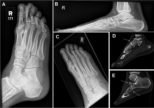
A 34 year-old male patient presented to Emergency department after twisting his right foot with tenderness around the calcaneal bone. Patient underwent an initial midfoot plain film (medial oblique and dorsoplanter) views. A cuboid bone fracture was initially missed on that admission report (Panel A). The Patient was sent home. Patient re-presented to Emergency department 5 days later with a persistent pain. Patient underwent further midfoot plain film (Lateral, dorsopanter and medial oblique) views (Panel B and C). Plain film report showed calcaneal anterior process fracture and cuboid bone fracture. Patient has been referred to Orthopaedic team and had subsequent CT scan (Panel D and E). CT scan confirmed the presence of calcaneal anterior process fracture and cuboid bone fracture. CT showed as well a navicular bone fracture could not be seen on initial plain films. No further imaging modalities were done to the patient. Patient has been followed by the orthopedic team with conservative management. This case clearly highlights the importance of all possible views for plain films and the need for subsequent imaging including CT scan if the clinical concern persists.

Figure: (A) Oblique view of right midfoot plain radiograph showing the missed cuboid bone fracture; (B) Lateral view of right midfoot plain radiograph showing the calcaneal anterior process fracture; (C) Dorsoplantar view of right midfoot plain radiograph showing the cuboid bone fracture; (D) CT sagittal view reconstructed image confirming right calcaneal anterior process fracture and cuboid bone fracture; (E) CT sagittal view reconstructed image showing right navicular bone fracture.
