Refractory Chylous Ascites after Living Donor Liver Transplantation Treated with Direct Percutaneous Embolization of Inguinal Lymphatic System: A Case Report
Article Information
Vinayak Nikam*, Kanchan Motwani, Nidhi Nair, Leena Tayshete, Gordhan Sangani, Preetha Joshi, Somnath Chattopadhyay
Department of HPB Surgery and Liver Transplantation, Kokilaben Dhirubhai Ambani Hospital, Andheri West, Mumbai, India
*Corresponding Author: Vinayak Nikam, Department of HPB Surgery and Liver Transplantation, Kokilaben Dhirubhai Ambani Hospital, Andheri West, Mumbai, India
Received: 17 June 2022; Accepted: 04 July 2022; Published: 26 July 2022
Citation: Vinayak Nikam, Kanchan Motwani, Nidhi Nair, Leena Tayshete, Gordhan Sangani, Preetha Joshi, Somnath Chattopadhyay. Refractory Chylous Ascites after Living Donor Liver Transplantation Treated with Direct Percutaneous Embolization of Inguinal Lymphatic System: A Case Report. Journal of Pediatrics, Perinatology and Child Health 6 (2022): 347-349.
Share at FacebookAbstract
Infection, trauma, cancer, or abdominal surgery can all induce chylous ascites. Chylous ascites (CA) following liver transplantation is extremely rare. The case we present here is of a patient who developed chylous ascites after a liver transplant and was treated with a combination of modalities before being resolved by lipiodol embolization of a lymphatic leak identified on fluorescent lymphangiography.
Keywords
Chylous Ascites, Liver Transplantation, Lymphatic Leak, Lymphangiogram, Lipiodol Embolization
Article Details
Abbreviations:
CA: Chylous Ascites; MCT: Medium chain triglyceride
1. Introduction
The extravasation of milky, triglyceride-rich lymphatic fluid within the peritoneal cavity is known as chylous ascites [1]. This is an uncommon complication of abdominal surgery, which is usually associated with trauma, cancer, or congenital malformations of the lymphatic system. The pathophysiological mechanisms governing this condition are related to a failure of the lymphatic system due to an obstruction caused by acquired or inherited factors [2]. Inadvertent injury to the lymphatic vessels during surgical procedures is one major contributor to CA occurring after liver transplants. The other mechanism is increased lymph production secondary to cirrhosis. The fact that more than 50% of the body’s lymphatic fluid originates in the gut and liver could be a contributing factor as well [3]. Less than 5% of recipients after liver transplantation report CA [4]. Because of the loss of vital lipids, proteins, water, vitamins, electrolytes, and immunoglobulins, chylous ascites can have serious and often fatal effects. Thus, the maintenance of adequate nutritional and immunological support while patients receive optimal therapy for CA is paramount, especially in pediatric patients and those on post-transplant immunosuppression. In our report, we would like to elaborate on a case of post-transplant chylous ascites which was managed with embolization of the injured lymphatic vessels.
2. Case Report
An 8-month old girl child, who was a known case of biliary atresia status post Kasai portoenterostomy procedure in October 2020, presented in April 2021 with chief complaints of jaundice, refusal of feeds, lethargy, irritability, and failure to thrive. She was diagnosed with end-stage liver disease and underwent a live donor liver transplant. On the 6th day postoperatively, she began to have chylous drain output and was started on a medium-chain triglyceride (MCT) diet. The drain fluid was sent for analysis, and it showed the triglyceride level to be 162 mg/dL. Drain output increased with time and was not resolving with the MCT diet alone. After 7 days of an exclusive MCT diet, there was still no improvement. She was then started on Total Parenteral Nutrition (TPN) and Inj Octreotide. Transient improvement was seen in the form of a decrease in the quantity of drain output, but its character remained chylous. Unfortunately, by postoperative day 21, her drain output began to increase again and was milky in colour.
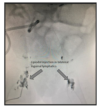
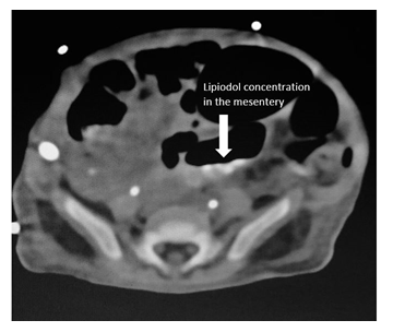
Day by day, her nutritional status deteriorated, and thus, an ultrasound and fluoroscopy guided lymphangiogram of the abdomen and pelvis was performed to further evaluate the source of chylous ascites. Under ultrasound guidance, intranodal inguinal lymph node injections of lipiodol with 25 gauge needles were conducted while the cephalad progression of the contrast was monitored. This demonstrated lymphatic duct filling all the way to the superior endplate of L2-L3. The site of lymphatic leaking was identified by contrast material pooling at L1-L2 (Figure 1). The embolization procedure was then completed, and a follow-up abdominal CT performed 24 hours later showed some lipiodol concentration on the mesenteric surface (Figure 2). Results of the successful embolization procedure were soon seen in the form of reduced chylous drainage and an improving overall nutritional status of the patient. The next 3-5 days after the lymphangiogram and embolization showed significantly decreased drain output, which led to drain removal on day 27 post-transplant. She was able to now accept full oral feeds and blood tests revealed normal graft function. The decision to discharge her was then made, and at the time of discharge she had normal liver function tests and no ascites. The patient is now 14 months post-transplant and is thriving. On latest follow up, her graft function remains good. She is found to be growing well and achieving all her developmental milestones as expected.
3. Discussion
Chylous Ascites after liver transplantation, though rare, is a known postoperative complication. Its incidence ranges from 0.6–4.7% in live liver transplant patients. The pathophysiology behind lymphatic fluid leaking into the abdominal cavity is multifactorial [5]. One mechanism is iatrogenic lymphatic vessel damage in the periportal and retrohepatic regions during the surgery and inadequate suturing of the injured vessels, leading to leakage of lymphatic fluid. Another mechanism of increased lymph leakage is an amplification in the lymph production occurring in the hepatic and splanchnic beds owing to postoperative adhesions and cirrhosis [4]. The criteria for diagnosing a patient with chylous ascites are: frank milky ascitic fluid when tapped and a fluid triglyceride value > 110 mg/dL [6]. Lymphangiograms are hailed to be the gold standard diagnostic tool to identify the source of the lymph leak in a patient with chylous ascites.
Treatment methodologies range from conservative dietary measures like the Medium Chain Triglyceride (MCT) diet and occasionally Total Parenteral Nutrition, to pharmacological means such as somatostatin analogues (Octreotide infusion). Fat-free enteral feeding with an MCT diet is generally the first line of management in cases of chylous ascites [1]. In most patients, these measures are sufficient to resolve the problem, but in cases where escalation of treatment is required, the addition of somatostatin analogues such as Octreotide has proved very effective. It is known to act upon gastric, pancreatic, and intestinal secretions as well as reduce splanchnic blood flow, resulting in decreased lymphatic fluid [7]. Intervention through radiological and surgical means is usually uncommon and only considered if all other conservative measures fail. When utilized, interventional radiological procedures like embolizations have a high success rate.
Instances of lymphatic leak identification during fluoroscopic lymphangiography are rare and are usually embolised in a CT-guided procedure using an N-butyl cyanoacrylate-lipiodol mixture. In some cases, the embolization is carried out solely with lipiodol with equally good results. During the extravasation of lipoidol, the ethiodized oil contrast agent, an inflammatory and granulomatous reaction occurs leading to sclerosis and obliteration of the point of leakage. It also has embolic qualities since it collects outside the leak source as well as within the lymphatics [8]. In cases unresponsive to treatment through non-surgical methods for over a month or in those showing an initial response but no recovery by the 2-month mark, surgical interventions in the form of ligation of leaking lymphatic vessels and shunt insertions may be considered [9]. In our patient, the embolization did not involve a CT-guided procedure but rather the lymphatic vessel leak was embolised using only lipiodol during the fluorescent lymphangiogram itself.
Declarations
Ethics approval committee name and consent to participate
- Institutional Ethics Committee, Kokilaben Dhirubhai Ambani Hospital, Mumbai, India.
- Father of recipient gave informed written consent for surgery and participation.
Consent for publication
Written and informed consent were taken from the father of recipient for the publication of this case report and the associated images.
- Availability of data and material- Not applicable.
- Competing interests- The authors declare that they have no competing interests.
- Funding- No grant support or any kind of assistance from other institute
- Authors' contributions
- Substantial contributions to the conception or design of the work; or the acquisition, analysis, or interpretation of data for the work -VN, KM, NN, LT, GS, PJ, SC.
- Drafting the work or revising it critically for important intellectual content- VN, KM, NN, LT, GS, PJ, SC.
- Final approval of the version to be published- VN, KM, NN, LT, GS, PJ, SC.
- Agreement to be accountable for all aspects of the work in ensuring that questions related to the accuracy or integrity of any part of the work are appropriately investigated and resolved- VN, KM, NN, LT, GS, PJ, SC.
Acknowledgements
The authors would like to thank Dr Diptiman Roy for IR procedure technical support, Dr Leena Tayshete and Mr. Rakesh Hire for the language editing help.
References
- Al-Busafi SA, Ghali P, Deschenes M, et al.
Chylous ascites: evaluation and management. ISRN Hepatol (2014): 240473.
- Lopez-Gutierrez JC, Tovar JA. Chylothorax and chylous ascites: management and pitfalls. Semin Pediatr Surg 23 (2014): 298-302.
- Mukerji AN, Tseng E, Karachristos A, et al. Chylous ascites after liver transplant: case report and review of literature. Exp Clin Transplant 4 (2013): 367-274.
- Yilmaz M, Akbulut S, Isik B, et al. Chylous ascites after liver transplantation: incidence and risk factors. Liver Transplant 18 (2012): 1046-1052.
- Dinc H, Oguz S, Sari A. A novel technique in the treatment of retroperitoneal lymphatic leakage: direct percutaneous embolization through the leakage pouch. Diagn Interv Radiol 21 (2015): 419-422.
- Riar S, Warshaw B, Ameral S. Chylous ascites complicating pediatric renal transplantation. Pediatr Nephrol 27 (2012): 1397-1399.
- Olivieri C, Nanni L, Masini L, et al. Successful management of congenital chylous ascites with early octreotide and total parenteral nutrition in a newborn. Case Reports (2012).
- Lee EW, Shin JH, Ko HK, et al. Lymphangiography to treat postoperative lymphatic leakage: A technical review. Korean Journal of Radiology 15 (2014): 724.
- Hwang JB, Choi S, Park WH. Resolution of refractory Chylous ascites after Kasai portoenterostomy using Octreotide. J Pediatr Surg 39 (2004): 1806.


