Questioning the Axiom: Is Hyperflexed Scaphoid a Common Occurrence in South Indian Population? - An Observational Study
Article Information
Mohamed Nazir ASHIK1, Maithreyi SETHU2*, Srinivasan RAJAPPA2
1Department of Orthopedics, Sri Ramachandra Institute of Higher Education and Research, No 1, Ramachandra Nagar, Sri Ramachandra Nagar, Chennai, 600116, Tamil Nadu, India
2Department of Hand Surgery, Sri Ramachandra Institute of Higher Education and Research, No 1, Ramachandra Nagar, Sri Ramachandra Nagar, Chennai, 600116, Tamil Nadu, India
*Corresponding Author: Maithreyi SETHU, Department of Hand Surgery, Sri Ramachandra Institute of Higher Education and Research, No 1, Ramachandra Nagar, Sri Ramachandra Nagar, Chennai, 600116, Tamil Nadu, India
Received: 28 June 2022; Accepted: 05 July 2022; Published: 12 July 2022
Citation: Mohamed Nazir ASHIK, Maithreyi SETHU, Srinivasan RAJAPPA. Questioning the Axiom: Is Hyperflexed Scaphoid a Common Occurrence in South Indian Population? - An Observational Study. Journal of Radiology and Clinical Imaging 5 (2022): 38-42.
Share at FacebookAbstract
Background: Scapholunate Angle SLA greater than 80° is one of the indicators of surgery for carpal instability. The normal SLA is between 30-60°. This is based on studies which were conducted several decades ago in the western part of the world. The SLA of Indian population has never been measured before. The knowledge of normal SLA in each population becomes imperative because it lays down the guidelines for surgical and non-surgical management of wrist fractures and instability in that population. In this study, we aim to measure the SLAs in asymptomatic volunteers of South Indian origin and compare the results with existing data.
Methods: An observational study was done in a tertiary care hospital in South India. Lateral radiographs of the wrist of asymptomatic volunteers were taken. The SLA was measured using software in the PACS system. The relation of SLA to gender, age, left and right sides and handedness was compared. These were compared to pre-existing global data available on SLAs.
Results: The SLA of 202 radiographs with 47 males and 54 females were studied.
• The average SLA was 51.33° and the range was between 29° and 74°.
• The average SLA of males was 51.8° and females 50.8°. There was no significant difference between the two.
• Similarly, there was no significant difference between the SLA of right and left sides in right handed and left handed subgroups.
• However, there was an increasing trend in SLA among females with age. But no similar pattern was seen among males.
• Interestingly, 39 out of the 202 radiographs (19.3%) had a SLA above 60°.
Conclusion: This study suggests the normal SLA in South Indians may be higher than the previously established reference values. This study serves as a pilot project and a reference for further studies on SLA.
Keywords
Scapholunate Angle (SLA); Scapholunate Dissociation (SLD); Carpal angle; Scaphoid; Lunate
Scapholunate Angle (SLA) articles; Scapholunate Dissociation (SLD) articles; Carpal angle articles; Scaphoid articles; Lunate articles
Scapholunate Angle articles Scapholunate Angle Research articles Scapholunate Angle review articles Scapholunate Angle PubMed articles Scapholunate Angle PubMed Central articles Scapholunate Angle 2023 articles Scapholunate Angle 2024 articles Scapholunate Angle Scopus articles Scapholunate Angle impact factor journals Scapholunate Angle Scopus journals Scapholunate Angle PubMed journals Scapholunate Angle medical journals Scapholunate Angle free journals Scapholunate Angle best journals Scapholunate Angle top journals Scapholunate Angle free medical journals Scapholunate Angle famous journals Scapholunate Angle Google Scholar indexed journals Scapholunate Dissociation articles Scapholunate Dissociation Research articles Scapholunate Dissociation review articles Scapholunate Dissociation PubMed articles Scapholunate Dissociation PubMed Central articles Scapholunate Dissociation 2023 articles Scapholunate Dissociation 2024 articles Scapholunate Dissociation Scopus articles Scapholunate Dissociation impact factor journals Scapholunate Dissociation Scopus journals Scapholunate Dissociation PubMed journals Scapholunate Dissociation medical journals Scapholunate Dissociation free journals Scapholunate Dissociation best journals Scapholunate Dissociation top journals Scapholunate Dissociation free medical journals Scapholunate Dissociation famous journals Scapholunate Dissociation Google Scholar indexed journals Carpal angle articles Carpal angle Research articles Carpal angle review articles Carpal angle PubMed articles Carpal angle PubMed Central articles Carpal angle 2023 articles Carpal angle 2024 articles Carpal angle Scopus articles Carpal angle impact factor journals Carpal angle Scopus journals Carpal angle PubMed journals Carpal angle medical journals Carpal angle free journals Carpal angle best journals Carpal angle top journals Carpal angle free medical journals Carpal angle famous journals Carpal angle Google Scholar indexed journals Scaphoid articles Scaphoid Research articles Scaphoid review articles Scaphoid PubMed articles Scaphoid PubMed Central articles Scaphoid 2023 articles Scaphoid 2024 articles Scaphoid Scopus articles Scaphoid impact factor journals Scaphoid Scopus journals Scaphoid PubMed journals Scaphoid medical journals Scaphoid free journals Scaphoid best journals Scaphoid top journals Scaphoid free medical journals Scaphoid famous journals Scaphoid Google Scholar indexed journals Lunate articles Lunate Research articles Lunate review articles Lunate PubMed articles Lunate PubMed Central articles Lunate 2023 articles Lunate 2024 articles Lunate Scopus articles Lunate impact factor journals Lunate Scopus journals Lunate PubMed journals Lunate medical journals Lunate free journals Lunate best journals Lunate top journals Lunate free medical journals Lunate famous journals Lunate Google Scholar indexed journals Anthropometry articles Anthropometry Research articles Anthropometry review articles Anthropometry PubMed articles Anthropometry PubMed Central articles Anthropometry 2023 articles Anthropometry 2024 articles Anthropometry Scopus articles Anthropometry impact factor journals Anthropometry Scopus journals Anthropometry PubMed journals Anthropometry medical journals Anthropometry free journals Anthropometry best journals Anthropometry top journals Anthropometry free medical journals Anthropometry famous journals Anthropometry Google Scholar indexed journals Hyperflexed Scaphoid articles Hyperflexed Scaphoid Research articles Hyperflexed Scaphoid review articles Hyperflexed Scaphoid PubMed articles Hyperflexed Scaphoid PubMed Central articles Hyperflexed Scaphoid 2023 articles Hyperflexed Scaphoid 2024 articles Hyperflexed Scaphoid Scopus articles Hyperflexed Scaphoid impact factor journals Hyperflexed Scaphoid Scopus journals Hyperflexed Scaphoid PubMed journals Hyperflexed Scaphoid medical journals Hyperflexed Scaphoid free journals Hyperflexed Scaphoid best journals Hyperflexed Scaphoid top journals Hyperflexed Scaphoid free medical journals Hyperflexed Scaphoid famous journals Hyperflexed Scaphoid Google Scholar indexed journals scapholunate ligament injuries articles scapholunate ligament injuries Research articles scapholunate ligament injuries review articles scapholunate ligament injuries PubMed articles scapholunate ligament injuries PubMed Central articles scapholunate ligament injuries 2023 articles scapholunate ligament injuries 2024 articles scapholunate ligament injuries Scopus articles scapholunate ligament injuries impact factor journals scapholunate ligament injuries Scopus journals scapholunate ligament injuries PubMed journals scapholunate ligament injuries medical journals scapholunate ligament injuries free journals scapholunate ligament injuries best journals scapholunate ligament injuries top journals scapholunate ligament injuries free medical journals scapholunate ligament injuries famous journals scapholunate ligament injuries Google Scholar indexed journals PAC articles PAC Research articles PAC review articles PAC PubMed articles PAC PubMed Central articles PAC 2023 articles PAC 2024 articles PAC Scopus articles PAC impact factor journals PAC Scopus journals PAC PubMed journals PAC medical journals PAC free journals PAC best journals PAC top journals PAC free medical journals PAC famous journals PAC Google Scholar indexed journals Scaphotrapeziotrapizoidal (STT) arthritis articles Scaphotrapeziotrapizoidal (STT) arthritis Research articles Scaphotrapeziotrapizoidal (STT) arthritis review articles Scaphotrapeziotrapizoidal (STT) arthritis PubMed articles Scaphotrapeziotrapizoidal (STT) arthritis PubMed Central articles Scaphotrapeziotrapizoidal (STT) arthritis 2023 articles Scaphotrapeziotrapizoidal (STT) arthritis 2024 articles Scaphotrapeziotrapizoidal (STT) arthritis Scopus articles Scaphotrapeziotrapizoidal (STT) arthritis impact factor journals Scaphotrapeziotrapizoidal (STT) arthritis Scopus journals Scaphotrapeziotrapizoidal (STT) arthritis PubMed journals Scaphotrapeziotrapizoidal (STT) arthritis medical journals Scaphotrapeziotrapizoidal (STT) arthritis free journals Scaphotrapeziotrapizoidal (STT) arthritis best journals Scaphotrapeziotrapizoidal (STT) arthritis top journals Scaphotrapeziotrapizoidal (STT) arthritis free medical journals Scaphotrapeziotrapizoidal (STT) arthritis famous journals Scaphotrapeziotrapizoidal (STT) arthritis Google Scholar indexed journals
Article Details
1. Introduction
Anthropometry is the measurement of living human individuals to understand the range of normal human physical variation [1]. To achieve accurate sex and age determination of normal values, every population should have its specific measurements [2]. The normal ranges of wrist measurements vary with race and ethnicity. The scapholunate angle plays an important role in the decision making of management of certain carpal bone and scapholunate ligament injuries [3]. With perilunate, wrist and hand injuries accounting for nearly 28% of all injuries [7], it is interesting to know what is the normal range in different ethnic groups.
The present study is the first to measure the Scapholunate Angles (SLA) of healthy South Indian population. The normal range of Scapholunate Angle (SLA) is between 30-60° [4-6]. This is based on studies which were conducted several decades ago in the western part of the world. The SLA of Indian population has never been measured before. The knowledge of normal SLA in each population becomes imperative because it lays down the guidelines for surgical and non-surgical management of wrist fractures and instability in that population. In this study, we aim to measure the SLAs in asymptomatic volunteers of South Indian origin and compare the results with existing data.
2. Methods
A descriptive observational study was conducted in a tertiary care centre in South India between January 2021 and July 2021. The aim was to measure the SLA in a normal, asymptomatic South Indian population and analyse any correlation with age, gender, between right and the left sides and handedness. Statistical analysis using average, standard deviation and range, significance of correlation of SLA with the different variables were calculated using the null hypothesis. Institutional ethical clearance was obtained before starting the study (CSP-MED /21/SEP/71/129). Inclusion criteria were patients above 20 years but less than 60 years with no specific complaints in upper limb, who were clinically normal and healthy volunteers. Exclusion criteria included children (in whom fusion of epiphysis is not yet complete), patients with acute or chronic wrist pain, recent or previous injury to the wrist, hand or forearm. Congenital conditions affecting the hand or wrist bones, infections and tumours of the hand and those with associated systemic condition causing hypercalcaemia or hypocalcaemia were also excluded from the study. Standard lateral radiographs of both wrists were taken.
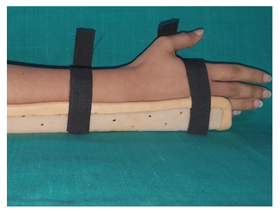
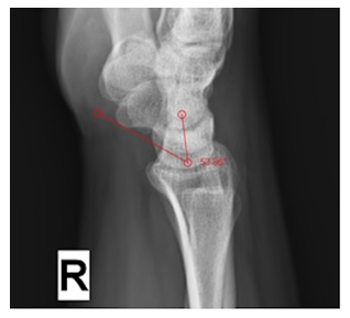
The scapholunate angle was measured using the software in PACS. These lateral radiographs were taken in standard Zero position with the arm adducted against the trunk, the elbow flexed 90° with the forearm in neutral rotation (no supination or pronation), and the wrist in neutral position (no radial or ulnar deviation, and no flexion or extension) [7]. We attained this using a below elbow thermoplastic splint as shown in Figure 1. It is similar to the Meyrueis’ technique, where a wooden board was used to maintain the forearm in a strict lateral position [8]. Rotational malalignment alters carpal bone measurements and by using this easy technique we attained the neutral wrist position [9]. Scapholunate angle was measured using the software in our PACS (Figure 2).

Figure 1: Thermoplastic splint aiding in keeping the wrist in neutral position during the radiograph.

Figure 2: Scapholunate Angle (SLA) subtended between the longitudinal lunate axis and the axis of the scaphoid attained by the tangential method.
|
Number |
Mean SLA |
SD |
P value |
|
|
Male |
94 |
50.80° |
9.29° |
0.432 |
|
Female |
108 |
51.76° |
8.39° |
|
|
SLA: Scapholunate Angle; SD: Standard Deviation |
||||
Table 1: Scapholunate Angle (SLA) comparison between males and females.
3. Results
The mean SLA of the 202 lateral radiographs was 51.3° with a standard deviation of 8.87°, ranging between 29° to 74° degrees. The average SLA of female was 51.8° and males 50.8° (Table 1). There was no significant difference between the two as indicated by the p-value. There was an increasing trend in the average SLA with age (Table 2).
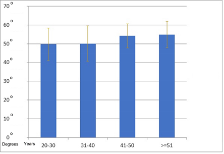
On further investigation it was found that women in the 20-30 years sub-group had a statistically significantly lower mean SLA when compared to the 40-50 years and above 50 years sub-group (Table 3). The SLA between intermediary age groups in the females showed a gradual increase but this was not statistically significant (Figure 3). In case of men, there was no statistical correlation between SLA and age.

Figure 3: Mean Scapholunate Angle (SLA) of females in different age group.
|
Age Group (Years) |
Number |
Mean SLA |
SD |
Standard error |
Range |
|
|
Min |
Max |
|||||
|
20-30 |
78 |
49.25° |
8.85° |
1 |
29.2 |
65.9 |
|
31-40 |
54 |
51.21° |
9.42° |
1.28 |
31.8 |
74.4 |
|
41-50 |
42 |
53.23° |
8.07° |
1.24 |
31.6 |
66.9 |
|
≥ 51 |
28 |
54.52° |
7.29° |
1.37 |
41.1 |
68.3 |
|
Total |
202 |
51.33° |
8.81° |
0.62 |
29.2 |
74.4 |
|
SLA: Scapholunate Angle; SD: Standard Deviation; Min: Minimum; Max: Maximum |
||||||
Table 2: Scapholunate Angle (SLA) distribution in different sub-groups of age.
|
Age Group (Years) |
Compared Age Group (Years) |
Significance (p value) |
|
|
Male |
20-30 |
31-40 |
0.08 |
|
41-50 |
0.38 |
||
|
≥51 |
0.09 |
||
|
31-40 |
20-30 |
0.08 |
|
|
41-50 |
0.53 |
||
|
≥51 |
0.86 |
||
|
41-50 |
20-30 |
0.38 |
|
|
31-40 |
0.53 |
||
|
≥51 |
0.47 |
||
|
≥51 |
20-30 |
0.91 |
|
|
31-40 |
0.86 |
||
|
41-50 |
0.47 |
||
|
Female |
20-30 |
31-40 |
0.87 |
|
41-50 |
0.04 |
||
|
≥51 |
0.04 |
||
|
31-40 |
20-30 |
0.87 |
|
|
41-50 |
0.05 |
||
|
≥51 |
0.05 |
||
|
41-50 |
20-30 |
0.49 |
|
|
31-40 |
0.53 |
||
|
≥51 |
0.77 |
||
|
≥51 |
20-30 |
0.04 |
|
|
31-40 |
0.05 |
||
|
41-50 |
0.77 |
Table 3: Comparison of Scapholunate Angle (SLA) with other age groups.
3.1 Variations among sides and dominance:
101 sets of right and left sided wrist radiographs were compared without taking gender or handedness into consideration. When SLA was compared, gender difference was not statistically significant (Table 4). Similarly, on comparing the handedness, there was no significant difference of SLA between dominant and non-dominant hands (Table 5).
|
Number |
Mean SLA |
SD |
P value |
|
|
Right |
101 |
50.81° |
8.88° |
P=0.245 |
|
Left |
101 |
51.85° |
8.76° |
|
|
SLA: Scapholunate Angle; SD: Standard Deviation |
||||
Table 4: Difference of Scapholunate Angle (SLA) between Right and Left side.
|
Number |
Mean SLA |
SD |
P value |
|
|
Dominant hand |
101 |
50.73° |
8.86° |
P=0.180 |
|
Non – Dominant hand |
101 |
51.93° |
8.76° |
|
|
SLA: Scapholunate Angle; SD: Standard Deviation |
||||
Table 5: Scapholunate Angle (SLA) and handedness.
4. Discussion
The scapholunate angle reflects the condition of scapholunate junction and is used to evaluate carpal bone alignment lateral radiographs [10]. On comparing the data of this study with other international literature available, the mean and range of SLA was in concurrence with study by Sarrafin et al [10], Larsen et al. [7], Jafari et al. [11]. But the range was much narrower in the data by Linsheid et al. [4], Schuid et al. [12] and Tang et al. when compared to our study (Table 6).
|
SLA Average |
SLA Range |
|
|
S.Indian |
51.3° |
29°-74° |
|
Linscheid et al. [4] |
46° |
30°-60° |
|
Sarrafian et al. [10] |
51° |
28°-101° |
|
Larsen et al. [7] |
50° |
- |
|
Schuind et al [12] |
45.8° (M) |
40.1°-51.5° |
|
48.4° (F) |
40.3°-56.5° |
|
|
Jafari et al. [11], Iran |
51.8° |
30°-80° |
|
Tang et al. [13], China |
54.7° |
48.2°-61.2° |
|
SLA: Scapholunate Angle; S: South |
||
Table 6: Comparison of Scapholunate Angle (SLA) among different Studies.
There was no significant difference in the average SLA between males and females in any of these studies (Table 7) but the range was much narrower in the data published by Schuhl et al. [8]. Again, there was no significant difference in the SLA based on handedness compared with other articles (Table 8).
|
SLA in Male (Mean ± SD) |
SLA in Female (Mean ± SD) |
|
|
S.Indian (This study) |
50.8° ± 9.29o |
51.76° ± 8.39° |
|
Schuind et al. [12] |
45.8° ± 5.7o |
48.4° ± 8.1° |
|
Schuhl et al. [8] |
25.52° - 43.32° |
29.2° - 49.9° |
|
Jafari et al. [11], Iran |
52.4° ± 8.5° |
51.5° ± 8.4° |
|
Tang et al. [13], China |
There was no significant difference in the SL angle (p=0.48), |
|
|
SLA: Scapholunate Angle; SD: Standard Deviation; S: South |
||
Table 7: Comparison of Scapholunate Angle (SLA) in genders among the different studies.
|
SLA in Dominant Hand |
SLA in Non-Dominant Hand |
|
|
S. Indian (This study) |
50.58° +52.8° |
- |
|
Schuind et al. [12] |
47.2° ± 6.5° |
46.6° ± 6.9° |
|
Schuhl et al. [8], Male |
32.8° ± 7.2° (23-51) |
36° ± 7.3° (2 2-48) |
|
Female |
39.5° ± 10.3o (17-55) |
39.3° ± 10.6° (11-84) |
|
SLA: Scapholunate Angle; S: South |
||
Table 8: Comparison of Scapholunate Angle (SLA) with handedness.
On comparing the SLA with age, there was no significance association in the study by Schuind et al. [12] and Jafari et al. [11], with the present study (Table 9). But an incidental finding in this study was that in case of females, the older women (>40years) had a significantly wider SLA than younger women (20-30years). This was not the same case with men. Another finding in this study was that 39 out of the 202 (19%) radiographs had SLA more than 60° which is considered as the normal upper limit which ranged from 61° to 74°. All of the 39 subjects were asymptomatic and none of them had an increased scapholunate gap. These interesting observations need further research and evaluation. It is beyond the scope of this paper to explore the reason behind it, but it definitely sparks a question of possibility if it is a common occurrence in many populations which has been missed so long.
|
SLA in Younger Age |
SLA in Older Age |
Significance |
|
|
S. Indian (This study) |
49.25° ± 8.85° |
54.52° ± 7.29° |
0.006 |
|
Schuind et al. [12] |
50.5° ± 6.2° |
43.7° ± 6.2° |
0.001 |
|
Jafari et al. [11], Iran |
51.1° ± 8.8° (20-40yrs) |
52.4o ± 8.0o (40-60 yrs.) |
NS |
|
Tang et al. [13], China |
SL angle did not change with patient’s age |
||
|
SLA: Scapholunate Angle; S: South; NS; Not significant |
|||
Table 9: Comparison of Scapholunate Angle (SLA) with age.
5. Limitation of the Study
Further research on Scapholunate Angle (SLA) is needed in many parts of the world to establish a wide consensus. The follow-up of the individuals especially those with a higher SLA might prove useful, to observe if they are more prone to Scaphotrapeziotrapizoidal (STT) arthritis in the later part of their lives. The significant increase of the SLA in female with age might also be a normal occurrence which has been unnoticed. But again, this needs to be addressed and confirmed with a multicentric study.
6. Conclusion
This research paper serves as a pilot study and as a reference for further studies on SLA angle in Indians. Though it is in concurrence with the previous articles on SLA pertaining to range, average and relation to age sex and handedness, it points out that further research and multicentric studies might be needed to redefine the normal range of SLA. The association of higher SLA with STT arthritis and the significant increase in SLA with age in women require long term cohort studies. The study might also influence designing of implants of carpal bones in the future.
Acknowledgements
Dr. T. Gayathri, Senior Lecturer in Statistics, Department of Allied Health Sciences, Sri Ramachandra Institute of higher Education and Research, Chennai, India
Conflict of Interest
The authors declare no conflicts of interest regarding the publication of this paper.
Funding
This research did not receive any specific grant from funding agencies in the public, commercial or not-for-profit sectors. Written informed consent was obtained from all the volunteers of this study. None of their identities were revealed in the study
Institutional Ethical Committee Approval
This study was approved by the Institutional Ethics Committee (CSP-MED /21/SEP/71/129).
Credit author statement
Dr. Mohamed Nazir ASHIK: Software, Investigation, Resources, Data Curation, Project administration.
Dr. Maithreyi SETHU: Methodology, Software, Validation, Formal analysis, Data Curation, Writing – Original, Writing - Review and Editing.
Dr. Srinivasan RAJAPPA: Conceptualization, Resources, Visualization, Supervision.
References
- Ali MHM. A normal data-base of posteroanterior radiographic measurements of the wrist in healthy Egyptians. Surgical and Radiologic Anatomy 31 (2009): 665-674.
- Franklin D, Freedman L, Milne N. Sexual dimorphism and discriminant function sexing in indigenous South African crania. HOMO- Journal of Comparative Human Biology 55 (2005): 213-228.
- Angermann P, Lohmann M. Injuries to the hand and wrist. A study of 50,272 injuries. J Hand Surg Br 18 (1993): 642-644.
- Linscheid RL, Dobyns JH, Beabout JW, et al. Traumatic instability of the wrist. The Journal of Bone & Joint Surgery 54 (1972): 1612-1632.
- Goldfarb CA, Yin Y, Gilula LA, et al. Wrist Fractures: What the Clinician Wants to Know. Radiology. 219 (2001):11-28.
- Gilula LA, Weeks PM. Post-traumatic Ligamentous Instabilities of the Wrist 1. Radiology 129 (1978): 641-651.
- Larsen CF, Stigsby B, Mathiesen FK, et al. Radiography of the wrist: A new device for standardized radiographs. Acta Radiologica 31(1990): 459-462.
- Schuhl J, Leroy B, Comtet J. cj. i tude rad.i.ologique de la mobiliti relative du scaphoide et du semi-lunaire radiological study of the mobility of the scapho-lunate joint. 4 (1985).
- Capo JT, Accousti K, Jacob G, et al. The effect of rotational malalignment on X-rays of the wrist. Journal of Hand Surgery: European Volume. 34 (2009): 166-172.
- Nakamura R, Hori M, Imamura T, et al. Method for measurement and evaluation of carpal bone angles. J Hand Surg Am 14 (1989): 412-416.
- Jafari D, Taheri H, Shariatzade H, et al. Radiographic indices in one hundred fifty normal Iranian wrists. Med J Islam Repub Iran 26 (2012): 132-139.
- Schuind F, Alemzadeh S, Stallenberg B, et al. Does the Normal Contralateral Wrist Provide the Best Reference for X-Ray Film Measurements of the Pathologic Wrist? J Hand Surg Am 21 (1996): 24-30.
- Tang CYK, Boris FKK, Man FMW, et al. A retrospective study of radiographic anatomy of wrist in Chinese population. E-Health Telecommunication Systems and Networks. 07 (2018): 57-65.
