Percutaneous Residual Stone Removal Post-lithotripsy During a Routine Nephrostogram: A Case Report and Interventional Implications
Article Information
Matthew Montanarella1*, Shannon Beardsley MD2
1Medical Student, Lake Erie College of Osteopathic Medicine, Bradenton, Florida, USA
2Department of Interventional Radiology, Baptist Medical Center, Jacksonville, Florida, USA
*Corresponding Author: Matthew Montanarella, Medical Student, Lake Erie College of Osteopathic Medicine, Bradenton, Florida, USA
Received: 23 January 2020; Accepted: 10 February 2020; Published: 18 February 2020
Citation: Matthew Montanarella, Shannon Beardsley MD. Percutaneous Residual Stone Removal Post-lithotripsy During a Routine Nephrostogram: A Case Report and Interventional Implications. Journal of Radiology and Clinical Imaging 3 (2020): 038-042.
Share at FacebookAbstract
Shock wave lithotripsy with percutaneous nephrolithotomy is an important treatment option for urolithiasis. Although this technique frequently removes the entirety of the stone, residual fragments of the stone may be unknowingly left within the urinary tract. When residual stones are visualized during post-procedure nephrostogram, interventional radiology can retrieve them percutaneously using the original access used by the surgeon for lithotripsy as is demonstrated in this case. Using a standard stone retrieval basket, or in this case report the basket component of an Arrow-Trerotola™ PTD® Percutaneous Thrombolytic Device, residual stones fragments of various sizes can be removed at the same session. If successful, the attendant risks and expense of additional urologic procedures to remove residual stones are avoided, thus demonstrating a potentially important role for interventional radiologists in the clinical management of these patients.
Keywords
Nephrostogram; Stone Fragment; Interventional Radiology; Percutaneous Nephrolithotomy
Nephrostogram articles, Stone Fragment articles, Interventional Radiology articles, Percutaneous Nephrolithotomy articles
Nephrostogram articles Nephrostogram Research articles Nephrostogram review articles Nephrostogram PubMed articles Nephrostogram PubMed Central articles Nephrostogram 2023 articles Nephrostogram 2024 articles Nephrostogram Scopus articles Nephrostogram impact factor journals Nephrostogram Scopus journals Nephrostogram PubMed journals Nephrostogram medical journals Nephrostogram free journals Nephrostogram best journals Nephrostogram top journals Nephrostogram free medical journals Nephrostogram famous journals Nephrostogram Google Scholar indexed journals Stone Fragment articles Stone Fragment Research articles Stone Fragment review articles Stone Fragment PubMed articles Stone Fragment PubMed Central articles Stone Fragment 2023 articles Stone Fragment 2024 articles Stone Fragment Scopus articles Stone Fragment impact factor journals Stone Fragment Scopus journals Stone Fragment PubMed journals Stone Fragment medical journals Stone Fragment free journals Stone Fragment best journals Stone Fragment top journals Stone Fragment free medical journals Stone Fragment famous journals Stone Fragment Google Scholar indexed journals Interventional Radiology articles Interventional Radiology Research articles Interventional Radiology review articles Interventional Radiology PubMed articles Interventional Radiology PubMed Central articles Interventional Radiology 2023 articles Interventional Radiology 2024 articles Interventional Radiology Scopus articles Interventional Radiology impact factor journals Interventional Radiology Scopus journals Interventional Radiology PubMed journals Interventional Radiology medical journals Interventional Radiology free journals Interventional Radiology best journals Interventional Radiology top journals Interventional Radiology free medical journals Interventional Radiology famous journals Interventional Radiology Google Scholar indexed journals Percutaneous Nephrolithotomy articles Percutaneous Nephrolithotomy Research articles Percutaneous Nephrolithotomy review articles Percutaneous Nephrolithotomy PubMed articles Percutaneous Nephrolithotomy PubMed Central articles Percutaneous Nephrolithotomy 2023 articles Percutaneous Nephrolithotomy 2024 articles Percutaneous Nephrolithotomy Scopus articles Percutaneous Nephrolithotomy impact factor journals Percutaneous Nephrolithotomy Scopus journals Percutaneous Nephrolithotomy PubMed journals Percutaneous Nephrolithotomy medical journals Percutaneous Nephrolithotomy free journals Percutaneous Nephrolithotomy best journals Percutaneous Nephrolithotomy top journals Percutaneous Nephrolithotomy free medical journals Percutaneous Nephrolithotomy famous journals Percutaneous Nephrolithotomy Google Scholar indexed journals Percutaneous Thrombolytic Device articles Percutaneous Thrombolytic Device Research articles Percutaneous Thrombolytic Device review articles Percutaneous Thrombolytic Device PubMed articles Percutaneous Thrombolytic Device PubMed Central articles Percutaneous Thrombolytic Device 2023 articles Percutaneous Thrombolytic Device 2024 articles Percutaneous Thrombolytic Device Scopus articles Percutaneous Thrombolytic Device impact factor journals Percutaneous Thrombolytic Device Scopus journals Percutaneous Thrombolytic Device PubMed journals Percutaneous Thrombolytic Device medical journals Percutaneous Thrombolytic Device free journals Percutaneous Thrombolytic Device best journals Percutaneous Thrombolytic Device top journals Percutaneous Thrombolytic Device free medical journals Percutaneous Thrombolytic Device famous journals Percutaneous Thrombolytic Device Google Scholar indexed journals shock wave lithotripsy articles shock wave lithotripsy Research articles shock wave lithotripsy review articles shock wave lithotripsy PubMed articles shock wave lithotripsy PubMed Central articles shock wave lithotripsy 2023 articles shock wave lithotripsy 2024 articles shock wave lithotripsy Scopus articles shock wave lithotripsy impact factor journals shock wave lithotripsy Scopus journals shock wave lithotripsy PubMed journals shock wave lithotripsy medical journals shock wave lithotripsy free journals shock wave lithotripsy best journals shock wave lithotripsy top journals shock wave lithotripsy free medical journals shock wave lithotripsy famous journals shock wave lithotripsy Google Scholar indexed journals percutaneous nephrolithotomy articles percutaneous nephrolithotomy Research articles percutaneous nephrolithotomy review articles percutaneous nephrolithotomy PubMed articles percutaneous nephrolithotomy PubMed Central articles percutaneous nephrolithotomy 2023 articles percutaneous nephrolithotomy 2024 articles percutaneous nephrolithotomy Scopus articles percutaneous nephrolithotomy impact factor journals percutaneous nephrolithotomy Scopus journals percutaneous nephrolithotomy PubMed journals percutaneous nephrolithotomy medical journals percutaneous nephrolithotomy free journals percutaneous nephrolithotomy best journals percutaneous nephrolithotomy top journals percutaneous nephrolithotomy free medical journals percutaneous nephrolithotomy famous journals percutaneous nephrolithotomy Google Scholar indexed journals Image articles Image Research articles Image review articles Image PubMed articles Image PubMed Central articles Image 2023 articles Image 2024 articles Image Scopus articles Image impact factor journals Image Scopus journals Image PubMed journals Image medical journals Image free journals Image best journals Image top journals Image free medical journals Image famous journals Image Google Scholar indexed journals Radiology articles Radiology Research articles Radiology review articles Radiology PubMed articles Radiology PubMed Central articles Radiology 2023 articles Radiology 2024 articles Radiology Scopus articles Radiology impact factor journals Radiology Scopus journals Radiology PubMed journals Radiology medical journals Radiology free journals Radiology best journals Radiology top journals Radiology free medical journals Radiology famous journals Radiology Google Scholar indexed journals Radiologists articles Radiologists Research articles Radiologists review articles Radiologists PubMed articles Radiologists PubMed Central articles Radiologists 2023 articles Radiologists 2024 articles Radiologists Scopus articles Radiologists impact factor journals Radiologists Scopus journals Radiologists PubMed journals Radiologists medical journals Radiologists free journals Radiologists best journals Radiologists top journals Radiologists free medical journals Radiologists famous journals Radiologists Google Scholar indexed journals
Article Details
1. Introduction
Procedural advances such as shock wave lithotripsy (SWL) and percutaneous nephrolithotomy (PCNL) have allowed practitioners to treat stones of varying sizes, shapes, and locations. Though the use of these procedures has aided in the treatment of urolithiasis, stones or stone fragments may be left behind after the procedure [1]. These stones were initially thought by some to be of little clinical significance. However, one-fifth of the patients with residual stones post PCNL, developed new clinically significant stones on the side affected [2]. If these stones do not receive treatment, half of them will cause some sort of stone related event, part of which will receive an additional surgical intervention [3,4]. We present a case of multiple residual stones discovered and removed during routine post-procedure nephrostogram.
2. Case Report
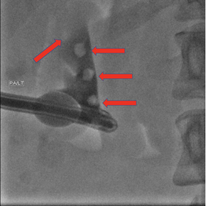
A 27-year-old female patient presented to interventional radiology on the morning of a scheduled operating room nephrolithotripsy for the treatment of a left partial staghorn calculus measuring approximately 2.5cm. The patient was brought into the angio suite for percutaneous access and placement of a 4-French Cobra glide catheter to the bladder. The patient was transferred to the operating room where the urologist performed tract dilation using an Amplatz renal dilator set followed by placement of a size 28-French sheath. This was followed by Cyberwand™ lithotripsy with percutaneous nephrolithotomy. At the end of the operating room procedure, a 20-French council-tip catheter was placed as a bridge to post-procedure nephrostogram. One week following the procedure, interventional radiology performed the nephrostogram. This was to evaluate for residual stones, extravasation, and ureteral patency before removal of the nephrostomy tube. Following a normal study, the tube would be removed. In our patients’ case, five stone fragments ranging from 4 to 6 mm were found in the pyelocalyceal system (Figure 1).

Figure 1: Fluoroscopic image demonstrating stone fragments (red arrows) within the pyelocalyceal system.
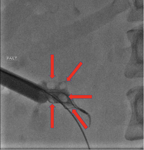
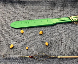
The nephrostomy tube was therefore exchanged for a 22-French Cook® Peel-Away® Sheath. Using the basket component of an Arrow-Trerotola™ PTD® Percutaneous Thrombolytic Device, each stone was removed percutaneously (Figure 2). Initial evaluation of the stone fragments demonstrated wide dispersion throughout the renal collecting system. We felt this may pose a challenge when attempting to remove them. However, when the sheath was introduced, the collecting system decompressed and the fragments all spontaneously moved to the central portion of the collecting system allowing for their easy retrieval. Figure 3 shows the removed stone fragments.

Figure 2: Intraoperative fluoroscopic image demonstrating the basket component of an Arrow-Trerotola™ PTD® Percutaneous Thrombolytic Device during removal of stone fragments (red arrows).

Figure 3: Intraoperative photograph of five stone fragments removed ranging from 4 to 6mm. Pictured below stones is the basket component of the Arrow-Trerotola™ PTD® Percutaneous Thrombolytic Device used in the procedure.
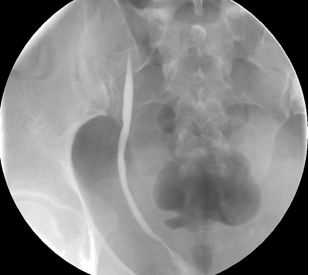
Post-procedure nephrostogram showed complete removal of residual stones and patency of the ureter (Figure 4). The tube was then removed.

Figure 4: Fluoroscopic image demonstrating a patent stone free distal ureter.
3. Discussion
Urolithiasis is a common condition that affects 10-15 percent of the world population [5]. Stones also have an estimated rate of reoccurrence at five years up to 50% [6]. The literature has shown that kidney stones are more common in men then they are in women, but recently that gap has narrowed and equalized amongst sexes [6]. In cases where other techniques such as lithotripsy or ureteroscopy fail, stones are larger than 20mm2, certain anatomical variants are present, or in cases of staghorn calculi, urology may elect to use PCNL as a method of stone removal [5]. Urologists often place a nephrostomy tube after lithotripsy to allow for a later nephrostogram by interventional radiology to ensure that there are no residual stones or obstruction before tube removal. Demonstrated in this paper is a case of a female patient that had multiple stone fragments inadvertently left behind after shock wave lithotripsy and percutaneous nephrolithotomy for a staghorn calculus. The fragments were successfully removed at the time of nephrostogram which eliminated the need for additional procedures. Without removal of the residual stone fragments, the nephrostomy tube would need to be left in place while the patient waits to see urology on a future visit. The urologist may then decide to place a double-J stent, remove the stone with a basket, or repeat nephrolithotripsy/nephrolithotomy, all of which would require additional scheduled procedures. Removal of residual stone fragments post-nephrostogram may reduce the need for these future surgical procedures and their associated risks as well as be a cost-effective means of treating the patient. In our case, we removed the fragments with the basket component of an Arrow-Trerotola™ PTD® Percutaneous Thrombolytic Device because it was readily available in the angio suite and we were familiar with its functionality. The device utilized was predicated on the standard ureteral basket device for stone retrieval. For subsequent cases of stone removal performed at our institution during post-procedure nephrostogram, we were successful with an 11mm standard urologic stone retrieval basket with a soft tip. We feel that a more widespread adoption of this technique by interventional radiologists may have a significant clinical and economic impact on care of these patients. Future studies demonstrating patient outcomes and procedural economics may be warranted.
Conflicts of Interest
The authors declared that there is no conflict of interest regarding the publication of this article.
References
- Candau C, Saussine C, Lang H, et al. Natural history of residual renal stone fragments after ESWL. EurUrol 37 (2000): 18-22.
- Osman MM, Alfano Y, Kamp S, et al. 5-year follow-up of patients with clinically insignificant residual fragments after extracorporeal shockwave lithotripsy. EurUrol 47 (2005): 860-864.
- Ozdedeli K, Cek M. Residual fragments after percutaneous nephrolithotomy. Balkan Med J 29 (2012): 230-235.
- Skolarikos A, Laguna MP, Alivizatos G. The role for active monitoring in urinary stones: a systematic review. J Endourol 24 (2010): 923-930.
- Ganpule A, Desai M. Fate of residual stones after percutaneous nephrolithotomy: A critical analysis. J Endourol 23 (2009): 399-403.
- Khan SR, Pearle MS, Robertson WG, et al. Kidney stones. Nat Rev Dis Primers 2 (2016): 16008.
