New Sign of Pneumothorax: Sharp Edge of Aorta on Prone Image
Article Information
Alan E. Oestreich*
Department of Radiology, Cincinnati Children’s Hospital Medical Center, Ohio, USA
*Corresponding Author: Dr. Alan E. Oestreich, Department of Radiology, Cincinnati Children’s Hospital Medical Center, Ohio, USA
Received: 20 October 2018; Accepted: 25 October 2018; Published: 30 October 2018
Citation: Alan E. Oestreich. New Sign of Pneumothorax: Sharp Edge of Aorta on Prone Image. Journal of Radiology and Clinical Imaging 1 (2018): 13-16
Share at FacebookAbstract
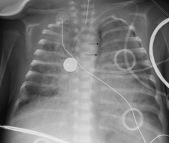
In the prone radiograph, a left pneumothorax may outline part of the upper descending aorta with a sharp interface (similar to the sharp edge of the lateral cardiac margin seen in supine radiographs). The sign is illustrated in a 2 month-old male infant with considerable bilateral lung disease.
Keywords
Pneumothorax, Descending aorta, Prone chest, Newly born, Sharp Edge sign
Pneumothorax, Descending aorta, Prone chest, Newly born, Sharp Edge sign
Pneumothorax articles Pneumothorax Research articles Pneumothorax review articles Pneumothorax PubMed articles Pneumothorax PubMed Central articles Pneumothorax 2023 articles Pneumothorax 2024 articles Pneumothorax Scopus articles Pneumothorax impact factor journals Pneumothorax Scopus journals Pneumothorax PubMed journals Pneumothorax medical journals Pneumothorax free journals Pneumothorax best journals Pneumothorax top journals Pneumothorax free medical journals Pneumothorax famous journals Pneumothorax Google Scholar indexed journals Descending aorta articles Descending aorta Research articles Descending aorta review articles Descending aorta PubMed articles Descending aorta PubMed Central articles Descending aorta 2023 articles Descending aorta 2024 articles Descending aorta Scopus articles Descending aorta impact factor journals Descending aorta Scopus journals Descending aorta PubMed journals Descending aorta medical journals Descending aorta free journals Descending aorta best journals Descending aorta top journals Descending aorta free medical journals Descending aorta famous journals Descending aorta Google Scholar indexed journals Prone chest articles Prone chest Research articles Prone chest review articles Prone chest PubMed articles Prone chest PubMed Central articles Prone chest 2023 articles Prone chest 2024 articles Prone chest Scopus articles Prone chest impact factor journals Prone chest Scopus journals Prone chest PubMed journals Prone chest medical journals Prone chest free journals Prone chest best journals Prone chest top journals Prone chest free medical journals Prone chest famous journals Prone chest Google Scholar indexed journals Newly born articles Newly born Research articles Newly born review articles Newly born PubMed articles Newly born PubMed Central articles Newly born 2023 articles Newly born 2024 articles Newly born Scopus articles Newly born impact factor journals Newly born Scopus journals Newly born PubMed journals Newly born medical journals Newly born free journals Newly born best journals Newly born top journals Newly born free medical journals Newly born famous journals Newly born Google Scholar indexed journals Sharp Edge sign articles Sharp Edge sign Research articles Sharp Edge sign review articles Sharp Edge sign PubMed articles Sharp Edge sign PubMed Central articles Sharp Edge sign 2023 articles Sharp Edge sign 2024 articles Sharp Edge sign Scopus articles Sharp Edge sign impact factor journals Sharp Edge sign Scopus journals Sharp Edge sign PubMed journals Sharp Edge sign medical journals Sharp Edge sign free journals Sharp Edge sign best journals Sharp Edge sign top journals Sharp Edge sign free medical journals Sharp Edge sign famous journals Sharp Edge sign Google Scholar indexed journals Prone Image articles Prone Image Research articles Prone Image review articles Prone Image PubMed articles Prone Image PubMed Central articles Prone Image 2023 articles Prone Image 2024 articles Prone Image Scopus articles Prone Image impact factor journals Prone Image Scopus journals Prone Image PubMed journals Prone Image medical journals Prone Image free journals Prone Image best journals Prone Image top journals Prone Image free medical journals Prone Image famous journals Prone Image Google Scholar indexed journals hypoxemic respiratory failure articles hypoxemic respiratory failure Research articles hypoxemic respiratory failure review articles hypoxemic respiratory failure PubMed articles hypoxemic respiratory failure PubMed Central articles hypoxemic respiratory failure 2023 articles hypoxemic respiratory failure 2024 articles hypoxemic respiratory failure Scopus articles hypoxemic respiratory failure impact factor journals hypoxemic respiratory failure Scopus journals hypoxemic respiratory failure PubMed journals hypoxemic respiratory failure medical journals hypoxemic respiratory failure free journals hypoxemic respiratory failure best journals hypoxemic respiratory failure top journals hypoxemic respiratory failure free medical journals hypoxemic respiratory failure famous journals hypoxemic respiratory failure Google Scholar indexed journals radiographic recognition articles radiographic recognition Research articles radiographic recognition review articles radiographic recognition PubMed articles radiographic recognition PubMed Central articles radiographic recognition 2023 articles radiographic recognition 2024 articles radiographic recognition Scopus articles radiographic recognition impact factor journals radiographic recognition Scopus journals radiographic recognition PubMed journals radiographic recognition medical journals radiographic recognition free journals radiographic recognition best journals radiographic recognition top journals radiographic recognition free medical journals radiographic recognition famous journals radiographic recognition Google Scholar indexed journals lateral decubitus image articles lateral decubitus image Research articles lateral decubitus image review articles lateral decubitus image PubMed articles lateral decubitus image PubMed Central articles lateral decubitus image 2023 articles lateral decubitus image 2024 articles lateral decubitus image Scopus articles lateral decubitus image impact factor journals lateral decubitus image Scopus journals lateral decubitus image PubMed journals lateral decubitus image medical journals lateral decubitus image free journals lateral decubitus image best journals lateral decubitus image top journals lateral decubitus image free medical journals lateral decubitus image famous journals lateral decubitus image Google Scholar indexed journals radiography. articles radiography. Research articles radiography. review articles radiography. PubMed articles radiography. PubMed Central articles radiography. 2023 articles radiography. 2024 articles radiography. Scopus articles radiography. impact factor journals radiography. Scopus journals radiography. PubMed journals radiography. medical journals radiography. free journals radiography. best journals radiography. top journals radiography. free medical journals radiography. famous journals radiography. Google Scholar indexed journals
Article Details
1. Introduction
Pneumothorax is a frequent respiratory tract complication in newborn infants. Radiographic perception of pneumothorax is well known for the supine image (sharp edge of the heart and the anterior hemidiaphragm); and on the upright image, it is found superiorly and laterally interfacing with lung. In the less frequent situation of a prone radiograph of an infant or older subject, recognition of a sharp edge of the upper descending aorta may permit a diagnosis of left pneumothorax.
2. Materials and Methods
A 2-month old male with left pneumothorax on prone radiographs is shown to illustrate the sharp edge of aorta as a sign of pneumothorax.
3. Results
A left pneumothorax is seen on Figure 1, where a sharp edge interface of air in the pleura abuts the sort tissue of the descending aorta. The subject died the following day. This is a sign apparently not previously reported - in my 50 years of radiology practice I have not recognized the sign in any other child.

Figure 1: Two month-old boy with bilateral lung disease; prone chest radiograph. The arrows indicate the sharp outline of the lateral upper descending aorta, a “sharp edge sign”. A stripe of lucency lateral to that interface represents responsible the posterior pneumothorax in this prone position. Further pneumothorax is noted at the lateral left hemithorax.
4. Discussion
Many clinical reasons for prone position are known, including open dysrhaphism in an infant and attempts to improve oxygenation in patients with hypoxemic respiratory failure [1]. Many signs of pneumothorax are well known for the supine and upright positions; analogous to the current sign is the sharp edge of the heart margin on the supine radiograph [2]. However, the radiographic recognition of pneumothorax in a prone position has not been well described.
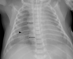
A sharper edge than normal appears when an x-ray beam is parallel to an interface between a soft tissue structure (such as the heart or the aorta) and pure air (as in pneumothorax), as opposed to less lucent usual lung air which contains also bronchi, blood vessels, and other lung contents. Such a sharp edge interface is the opposite of the silhouette sign in which an expected lung-soft tissue interface is lost because of soft tissue interfacing with soft tissue. The sharp edge of the right heart (and thymus) border from an anterior pneumothorax on a supine chest is illustrated by Figure 2 and is explained by diagrams in reference [2].
A lung that is diseased tends not to collapse completely when pneumothorax is present. In supine position a pneumothorax rises anteriorly.

Figure 2: Two day-old boy following surfactant administration for surfactant deficiency disease with increased respiratory distress and oxygen desaturation; supine chest radiograph. A right anterior pneumothorax in indicated by the sharp edge of the heart and thymus (arrows). The arrowhead indicates the less sharp lateral margin of the pneumothorax. Note that the left heart margin is far less sharp than the right. The right anterior hemidiaphragm is also more sharply seen than the left, due to the anterior pneumothorax. The pneumothorax was confirmed by a left side down lateral decubitus image.
By analogy to the anterior pneumothorax sharp edge against the heart (and anterior hemidiaphragm) in supine position, a sharp edge of the lateral margin of the upper descending aorta from a posterior pneumothorax in the prone position (as in Figure 1) is similarly explained by purely air of a pneumothorax interfacing with soft tissue. As with anterior pneumothorax in supine position, the lateral margin of the pneumothorax may be less sharply defined, being a laterally tapered edge of the pneumothorax.
Our subject shown in Figure 1 had bilateral lung disease and died the day after the demonstrated prone image was obtained; no autopsy was obtained. His chest radiograph serves to illustrate a vagary of the sharp edge sign and we offer it as a new sign of posterior pneumothorax potentially seen on prone patient radiography.
Conflict of interest
None
References
- Kopterides P, Siemos II, Armagannidis A. Prone Position in Hypoxic Respiratory Failure: Meta-analysis of randomized controlled trials. Journal of Critical Care 24 (2009): 89-100.
- Oestreich AE. The Sharp Edge; A Frequent Sign in Neonatal Pneumothorax. Journal of Medical Imaging 1 (1987): 283-286.
