Mixed Small Cell Carcinoma and Adenocarcinoma of the Esophagus: A Case Report
Article Information
Tasneem Dawood1*, Muhammad Nauman Zahir2, Bilal Mazhar Qureshi1, Zeeshan Uddin1, Yasmin Abdul Rashid1, Adnan Abdul Jabbar2
1Aga Khan University Hospital, Karachi, Pakistan
2Ziauddin University Hospital, Karachi, Pakistan
*Corresponding Author: Tasneem Dawood, Aga Khan University Hospital, Karachi, Pakistan.
Received: 01 September 2022; Accepted: 21 September 2022; Published: 16 November 2022
Citation: Tasneem Dawood, Muhammad Nauman Zahir, Bilal Mazhar Qureshi, Zeeshan Uddin, Yasmin Abdul Rashid, Adnan Abdul Jabbar. Mixed Small Cell Carcinoma and Adenocarcinoma of the Esophagus: A Case Report. Archives of Clinical and Medical Case Reports 6 (2022): 734-737.
Share at FacebookAbstract
Background: Small cell carcinoma is an aggressive malignant tumor characterized by small-sized cells with scant cytoplasm, nuclei with finely granular chromatin and absent nucleoli, and a high mitotic count. The most common site is the lung which comprises 10-15 percent of all lung cancers. Rare sites of small cell carcinoma include the uterus, cervix, prostate, larynx, sweat glands, and gastrointestinal tract. No matter the site, it is an aggressive disease and is usually associated with a poor prognosis.
Case Summary: We report the case of a 31-year-old gentleman, diagnosed with mixed small cell carcinoma and adenocarcinoma of the esophagus. He presented to our clinic with complaints of progressive dysphagia, cough, and weight loss for the last 6 months. An initial endoscopic biopsy was done, it was suspicious of mixed small cell carcinoma and adenocarcinoma of the esophagus which was reconfirmed by our histopathologist. MRI brain was negative. He was treated aggressively with Cisplatin and Etoposide-based concurrent chemo-radiation. He received 4 cycles of chemotherapy and 28 fractions of radiation sandwiched between cycles 2 and 4 of chemotherapy. He has been on surveillance since April 2019 and his most recent scan and upper GI endoscopy from May 2019 confirm that the disease is in remission.
Conclusion: Mixed Small cell carcinoma and adenocarcinoma of the esophagus is an extremely unique, aggressive, and swiftly progressive disease, prone to distant metastasis and poor prognosis if left untreated. Treatment should be aggressive and commenced as soon as possible. Multi-modality management should be considered the current standard of care, to achieve both local and distant disease control.
Keywords
Chemoradiation; Multi-Modality; Small Cell
Chemoradiation articles; Multi-Modality articles; Small Cell articles
Chemoradiation articles Chemoradiation Research articles Chemoradiation review articles Chemoradiation PubMed articles Chemoradiation PubMed Central articles Chemoradiation 2023 articles Chemoradiation 2024 articles Chemoradiation Scopus articles Chemoradiation impact factor journals Chemoradiation Scopus journals Chemoradiation PubMed journals Chemoradiation medical journals Chemoradiation free journals Chemoradiation best journals Chemoradiation top journals Chemoradiation free medical journals Chemoradiation famous journals Chemoradiation Google Scholar indexed journals COVID-19 articles COVID-19 Research articles COVID-19 review articles COVID-19 PubMed articles COVID-19 PubMed Central articles COVID-19 2023 articles COVID-19 2024 articles COVID-19 Scopus articles COVID-19 impact factor journals COVID-19 Scopus journals COVID-19 PubMed journals COVID-19 medical journals COVID-19 free journals COVID-19 best journals COVID-19 top journals COVID-19 free medical journals COVID-19 famous journals COVID-19 Google Scholar indexed journals Multi-Modality articles Multi-Modality Research articles Multi-Modality review articles Multi-Modality PubMed articles Multi-Modality PubMed Central articles Multi-Modality 2023 articles Multi-Modality 2024 articles Multi-Modality Scopus articles Multi-Modality impact factor journals Multi-Modality Scopus journals Multi-Modality PubMed journals Multi-Modality medical journals Multi-Modality free journals Multi-Modality best journals Multi-Modality top journals Multi-Modality free medical journals Multi-Modality famous journals Multi-Modality Google Scholar indexed journals Ultrasound articles Ultrasound Research articles Ultrasound review articles Ultrasound PubMed articles Ultrasound PubMed Central articles Ultrasound 2023 articles Ultrasound 2024 articles Ultrasound Scopus articles Ultrasound impact factor journals Ultrasound Scopus journals Ultrasound PubMed journals Ultrasound medical journals Ultrasound free journals Ultrasound best journals Ultrasound top journals Ultrasound free medical journals Ultrasound famous journals Ultrasound Google Scholar indexed journals Small Cell articles Small Cell Research articles Small Cell review articles Small Cell PubMed articles Small Cell PubMed Central articles Small Cell 2023 articles Small Cell 2024 articles Small Cell Scopus articles Small Cell impact factor journals Small Cell Scopus journals Small Cell PubMed journals Small Cell medical journals Small Cell free journals Small Cell best journals Small Cell top journals Small Cell free medical journals Small Cell famous journals Small Cell Google Scholar indexed journals Radiotherapy articles Radiotherapy Research articles Radiotherapy review articles Radiotherapy PubMed articles Radiotherapy PubMed Central articles Radiotherapy 2023 articles Radiotherapy 2024 articles Radiotherapy Scopus articles Radiotherapy impact factor journals Radiotherapy Scopus journals Radiotherapy PubMed journals Radiotherapy medical journals Radiotherapy free journals Radiotherapy best journals Radiotherapy top journals Radiotherapy free medical journals Radiotherapy famous journals Radiotherapy Google Scholar indexed journals CT articles CT Research articles CT review articles CT PubMed articles CT PubMed Central articles CT 2023 articles CT 2024 articles CT Scopus articles CT impact factor journals CT Scopus journals CT PubMed journals CT medical journals CT free journals CT best journals CT top journals CT free medical journals CT famous journals CT Google Scholar indexed journals Prognosis articles Prognosis Research articles Prognosis review articles Prognosis PubMed articles Prognosis PubMed Central articles Prognosis 2023 articles Prognosis 2024 articles Prognosis Scopus articles Prognosis impact factor journals Prognosis Scopus journals Prognosis PubMed journals Prognosis medical journals Prognosis free journals Prognosis best journals Prognosis top journals Prognosis free medical journals Prognosis famous journals Prognosis Google Scholar indexed journals Seasonality articles Seasonality Research articles Seasonality review articles Seasonality PubMed articles Seasonality PubMed Central articles Seasonality 2023 articles Seasonality 2024 articles Seasonality Scopus articles Seasonality impact factor journals Seasonality Scopus journals Seasonality PubMed journals Seasonality medical journals Seasonality free journals Seasonality best journals Seasonality top journals Seasonality free medical journals Seasonality famous journals Seasonality Google Scholar indexed journals Teledentistry articles Teledentistry Research articles Teledentistry review articles Teledentistry PubMed articles Teledentistry PubMed Central articles Teledentistry 2023 articles Teledentistry 2024 articles Teledentistry Scopus articles Teledentistry impact factor journals Teledentistry Scopus journals Teledentistry PubMed journals Teledentistry medical journals Teledentistry free journals Teledentistry best journals Teledentistry top journals Teledentistry free medical journals Teledentistry famous journals Teledentistry Google Scholar indexed journals
Article Details
1. Introduction
Esophageal carcinoma is the eighth most common cancer worldwide. It is mainly characterized by two histological subtypes: namely squamous cell carcinoma and adenocarcinoma. Small cell carcinoma of the esophagus is a rare histological subtype accounting for only 1-2.8% of all esophageal cancers [1]. It is a highly aggressive disease with a high risk of distant metastasis and a poor prognosis. The median survival is usually less than a year. The recurrence rate is very high even in patients with early-stage disease [1]. Mixed small cell and adenocarcinoma of the esophagus are exceedingly rare [2]. Small cell carcinoma also known as oat cell carcinoma is a neuroendocrine carcinoma that exhibits aggressive behavior, rapid proliferation, and high rates of distant metastasis. It is frequently associated with paraneoplastic syndromes such as hypercalcemia, Lambert Eaton syndrome, SIADH, sub-acute cerebellar degeneration, limbic encephalopathy, and acromegaly [3]. The site of small cell carcinoma is usually the lung. Extrapulmonary small cell carcinoma comprises less than 5% of cases and is rarely seen in sites such as the salivary glands, larynx, pharynx, cervix, prostate, breast, and gastrointestinal tract [4]. A combined modality approach is usually used for treatment including chemotherapy and radiation based on studies on small cell carcinoma of the lung [5]. Small Cell carcinoma of the lung is divided into limited stage and extensive stage. The limited stage is defined as disease that is limited to the ipsilateral hemi thorax and regional lymph nodes and can be encompassed in a safe radiotherapy field. Extensive stage (ES) disease is defined as disease with distant metastases, malignant pericardial or pleural effusions, and/or contralateral supraclavicular and contralateral hilar lymph node involvement [6]. For Limited stage disease of the lung, some patients are candidates for lobectomy with mediastinal sampling or dissection. If surgery demonstrates mediastinal nodal involvement, adjuvant chemoradiation is indicated. If they are not surgical candidates, stereotactic ablative body therapy (SABR) may be offered. Adjuvant cisplatin-based systemic therapy typically follows both local approaches. The rest of the limited-stage patients are usually treated with concurrent chemotherapy and radiation. The chemotherapeutic agents used are cisplatin and etoposide are the current standard of care [6]. The mainstay of therapy for ES SCLC is systemic therapy. It is a fundamental part of management, as these tumors have a high proliferative index and respond well to aggressive treatment. Radiotherapy alone as a treatment modality is usually not helpful [5]. Here we report the case of a young male diagnosed with mixed small cell carcinoma and adenocarcinoma of the esophagus and how he was managed using a multimodality approach.
2. Case Report
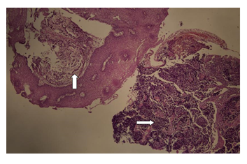
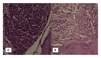
A 31-year-old young gentleman, who had a history of occasional cigarette smoking with no other significant past medical history, presented to the clinic in January 2019 with complaints of dysphagia along with chest tightness, epigastric burning, and loss of appetite and weight loss over the last 4-5 months. His Upper GI endoscopy was done outside our institute in December 2018 which revealed a 3 cm ulcerated semi-circumferential mass extending from 38 cm to 41 cm to the gastroesophageal junction along with mild pan gastritis. The biopsy of the esophageal mass was taken. The biopsy was reviewed again at our institute, and it showed esophageal mucosal fragments involved by an invasive tumor composed of partly crushed small round to oval cells arranged in sheets and clusters. Individual cells show scant cytoplasm and nuclear hyperchromasia. Focally the tumor cells are forming well-formed glands. Immunohistochemical stains revealed CK7, CD56, and CDX 2 positive with a high proliferative index of 80%. Features were consistent with mixed small cell carcinoma and adenocarcinoma of the esophagus. CT scan of the chest and liver was done outside our institute which showed an asymmetrical soft tissue mass with ulceration causing wall thickening of the distal esophagus and involving the GEJ along with prominent gastro hepatic lymph nodes. PET CT scan was also done which revealed limited stage of the disease. MRI brain was unremarkable. His Hepatitis B/C and HIV test were all negative and 24-hour creatinine clearance and including all other baseline labs were fine. This case was discussed in the multidisciplinary tumor board meeting. It was decided to treat the patient as limited stage small cell carcinoma of the lung with multimodality treatment. Chemotherapy was started, Cisplatin 80mg/m2 day 1 and Etoposide 100mg/m2 day 1-3 x 4 cycles given every 3 weeks. The first cycle was given before radiation. A feeding tube was placed in February 2019 before radiation was started. The patient was later started on definitive concurrent chemoradiation again with Cisplatin 80mg/m2 day 1 and Etoposide 100mg/m2 day 1-3 x given every 3 weeks with 50.4 Gy in 28 fractions of radiation with the remaining chemotherapy cycles. 4 cycles of chemotherapy were completed in April 2019 and radiation ended in March 2019. No prophylactic cranial irradiation was given. CT scan was done after completion of treatment in May 2019 which showed redemonstration of mild thickening in the region of the gastro-esophageal junction extending to involve the cardia of the stomach, which appeared grossly unchanged from prior examination. No evidence of distant metastasis. Upper GI endoscopy was also repeated after completion of treatment, which showed no evidence of disease. His feeding tube was then removed. He has been on surveillance since then. The scans repeated in August 2019 and January 2020, were unremarkable. He was followed up every 3 months for history and physical exam with baseline labs and CT scans were repeated every 6 months. His latest scan was done in June 2020 which also showed no evidence of disease.

Figure 1: Esophageal biopsy showing mixed small cell carcinoma (→) and adenocarcinoma (↑). Normal stratified squamous epithelium of esophagus is present in the center (H&E, 4X).

Figure 2: Esophageal biopsy showing cell carcinoma component (A), composed of sheets of undifferentiated small cells with hyperchromatic nuclei and nuclear molding. Adenocarcinoma component (B) is composed of complex glands lined by neoplastic cells and lumina showing secretion (H&E, 20X).

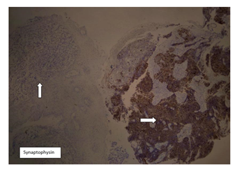
Figure 3: Immunohistochemical stain synaptophysin on the esophageal biopsy showing positivity in small cell carcinoma component (→), consistent with neuroendocrine differentiation. The adenocarcinoma component (↑) is negative for this marker. (4X).
3. Discussion
Squamous cell carcinoma (SCC) and adenocarcinoma account for 95 percent of histological subtypes of esophageal cancer. Primary small cell carcinoma of the esophagus is a rare disease accounting for 1-2.8 of esophageal cancer. It is a very aggressive disease and is notorious for early distant metastasis and poor prognosis. Small cell carcinoma of the esophagus can cause early dissemination and usually has a median survival of less than a year. It even has a high recurrence rate [1]. Small cell carcinoma is histologically thought to develop from neuroendocrine Kulchitsky cells and is composed of sheets of small, round to spindled cells with dark nuclei, scarce cytoplasm, and fine, granular (“salt and pepper”) nuclear chromatin with indefinite nucleoli [7]. Small cell carcinoma accounts for 14-15% of lung cancers [8]. Extra pulmonary small cell carcinomas (ESCCs) are very rare; these tumors have been described most frequently in the urinary bladder, prostate, esophagus, stomach, colon and rectum, gallbladder, larynx, salivary glands, cervix, and skin. In addition, small cell carcinoma will occasionally present with metastatic disease, and a primary site cannot be identified (small cell carcinoma of unknown primary) [9]. Primary small cell carcinoma of the esophagus has similar findings on endoscopy and radiology as the other subtypes. But usually presents with rapid dysphagia and weight loss in the early days. This subtype is mostly seen in men with a male-to-female ratio reported as 2: 1. It is usually observed between the fourth and the seventh decades. Major symptoms include progressive dysphagia, retrosternal pain, cough, and rapid weight loss. Rarely, hoarseness and upper gastrointestinal tract bleeding have also been reported as the primary symptoms. It is usually seen in the distal or middle esophagus. Sites of distant spread include liver, lung, and bones [5]. There are 2 viewpoints regarding the pathogenesis of primary small cell carcinoma of the esophagus, one is that it arises from neuroendocrine cells of the submucosal gland or stratum basal, and the major precursor from the APUD cells is the amine precursor uptake decarboxylase cells. Since these cells are abundant in the distal esophagus, the lesion usually arises in the middle or distal esophagus. The second is that the biphasic neoplasm originates from pluripotential stem cells of the endoderm that can be partially differentiated into the squamous cell, neuroendocrine cell, or glandular cell because of the stimulation of different carcinogenic agents [5]. We could not find any cases reported yet, regarding mixed small cell carcinoma and adenocarcinoma of the Esophagus. Most of the cases reported include mixed small cell and squamous cell carcinoma of the esophagus [11,12]. No standard of care of treatment has yet been established because of the rarity of the disease. The significance of surgery is still controversial [5]. Some reports have stressed that surgery should be avoided for patients with advanced disease [13]. Conversely, some authors think surgery remains the primary method in patients with localized disease [14]. In recent reports, regimens including cisplatin and etoposide have achieved better response and radiotherapy is also effective [14]. Several cases suggested that the patients were treated with surgical resection, radiotherapy, and chemotherapy in combination may result in survival benefits [15]. Nonetheless, individualized treatment should be considered for all patients, based on clinical features, pathologic diagnosis, the grading and staging classification, patient characteristics, logistics, and availability of surgical expertise as incomplete surgery or recovery might delay chemoradiation which has proven to be effective for locoregional and distant control of disease [16].
4. Conclusion
Primary mixed small cell carcinoma and adenocarcinoma of the esophagus is an extremely rare disease with high risk of early dissemination and poor prognosis if left untreated. It should be dealt with aggressively and treatment decisions should be made early. These cases should be discussed in a multidisciplinary meeting and a plan should be made personalized to each patient. Treatment should be aggressive and started early. Multi-modality management should be considered the current standard of care in such patients to help achieve, both local and distant disease control. These patients need a close follow-up post-treatment because of the aggressive nature of the disease and the high risk of recurrence.
Conflicts of Interest
The authors have no conflicts of interest to declare. All co-authors have seen and agree with the contents of the manuscript and there is no financial interest to report. We certify that the submission is original work and is not under review at any other publication.
References
- Chen WW, Wang F, Zhang DS, et al. Primary small cell carcinoma of the esophagus: clinicopathological study of 44 cases. BMC cancer 14 (2014): 222.
- Schwartz CJ, Hickman R, Zhang X, et al. Primary Esophageal Mixed Sarcomatoid and Small Cell Neuroendocrine Carcinoma With Brain Metastasis: A Challenging Diagnosis on Biopsy. International Journal of Surgical Pathology 27 (2018): 84-88.
- Kanaji N, Watanabe N, Kita N, et al. Paraneoplastic syndromes associated with lung cancer. World journal of clinical oncology 5 (2014): 197-223.
- Gennatas S, Noble J, Stanway S, et al. Patterns of relapse in extrapulmonary small cell carcinoma: retrospective analysis of outcomes from two cancer centres. BMJ Open 5 (2015): e006440.
- Yekeler E, Koca T, Vural S. A Rare Cause of the Cough: Primary Small Cell Carcinoma of Esophagus—Case Report. Case Reports in Medicine 2012 (2012): 870783.
- Wang S, Zimmermann S, Parikh K, et al. Current Diagnosis and Management of Small-Cell Lung Cancer. Mayo Clinic proceedings 94 (2019): 1599-622.
- Zakowski MF. Pathology of small cell carcinoma of the lung. Seminars in oncology 30 (2003): 3-8.
- Dorantes-Heredia R, Ruiz-Morales JM, Cano-García F. Histopathological transformation to small-cell lung carcinoma in non-small cell lung carcinoma tumors. Transl Lung Cancer Res 5 (2016): 401-412.
- Wong YNS, Jack RH, Mak V, et al. The epidemiology and survival of extrapulmonary small cell carcinoma in South East England, 1970-2004. BMC cancer 9 (2009): 209.
- Glatzer M, Schmid S, Radovic M, et al. The role of radiation therapy in the management of small cell lung cancer. Breathe (Sheff) 13 (2017): e87-e94.
- Muguruma K, Ohira M, Tanaka H, et al. Long-term Survival of Advanced Small Cell Carcinoma of the Esophagus after Resection: A Case Report. Anticancer Research 33 (2013): 595.
- Goh N, Yeo DXW, Amitbhai SK, et al. A rare case of bipartite combined tumour of the oesophagus. World Journal of Surgical Oncology 17 (2019): 79.
- Isolauri J, Mattila J, Kallioniemi OP. Primary undifferentiated small cell carcinoma of the esophagus: Clinicopathological and flow cytometric evaluation of eight cases. Journal of Surgical Oncology 46 (1991): 174-177.
- Yachida S, Matsushita K, Usuki H, et al. Long-term survival after resection for small cell carcinoma of the esophagus. The Annals of thoracic surgery 72 (2001): 596-597.
- Tobari S, Ikeda Y, Kurihara H, et al. Effective treatment with chemotherapy and surgery for advanced small cell carcinoma of the esophagus. Hepato-gastroenterology 51 (2004): 1027-1029.
- Hudson E, Powell J, Mukherjee S, et al. Small cell oesophageal carcinoma: an institutional experience and review of the literature. Br J Cancer 96 (2007): 708-711.
