Minor Salivary Glands and ‘Tubarial Glands’-Anatomy, Physiology, and Pathology Relevant to Radiology
Article Information
Sabujan Sainudeen1*, Asmi Sabujan2
1Department of Radiology, Thangam Hospital, Palakkad, Kerala, India
2Department of Radiology, Malabar Scans and Research centre, Tirur, Kerala, India
*Corresponding Author: Dr. Sabujan Sainudeen, Department of Radiology, Thangam Hospital, Palakkad, Kerala, India
Received: 24 December 2020; Accepted: 19 January 2021; Published: 26 January 2021
Citation: Sabujan Sainudeen, Asmi Sabujan. Minor Salivary Glands and ‘Tubarial Glands’-Anatomy, Physiology, and Pathology Relevant to Radiology. Journal of Radiology and Clinical Imaging 4 (2021): 001-014.
Share at FacebookAbstract
Tubarial glands or tubarial salivary glands are recently reported as a pair of macroscopic salivary glands in the nasopharynx. The remote location of the glands, the rarity of major pathologies involved, and non recognized functional significance might have been the reason for the non-inclusion before. There are about 500-1000 minor salivary glands in the body, and most of them are located in the oral cavity or oropharynx. They are small and embedded in the aero-digestive tract entrance of the head and neck region. Minor salivary glands located in the nasopharynx are relatively less in number and the major pathologies associated with these glands are rare. This review is on the overall current literature of the minor salivary glands and tubarial glands-the new entity in question-along with their potential pathologies. Nasopharyngeal glandular origin diseases were reported in general as case reports or as small series. This brief review is meant to open up interest in these structures, their pathologies and encourage further characterization of diseases of the nasopharynx especially the diseases of salivary gland origin.
Keywords
Salivary glands; Tubarial glands; Minor salivary glands; Tubarial salivary glands; Nasopharynx; Torus tubaris; Fossa of Rosenmüller
Salivary glands articles; Tubarial glands articles; Minor salivary glands articles; Tubarial salivary glands articles; Nasopharynx articles; Torus tubaris articles; Fossa of Rosenm?ller articles
Salivary glands articles Salivary glands Research articles Salivary glands review articles Salivary glands PubMed articles Salivary glands PubMed Central articles Salivary glands 2023 articles Salivary glands 2024 articles Salivary glands Scopus articles Salivary glands impact factor journals Salivary glands Scopus journals Salivary glands PubMed journals Salivary glands medical journals Salivary glands free journals Salivary glands best journals Salivary glands top journals Salivary glands free medical journals Salivary glands famous journals Salivary glands Google Scholar indexed journals Tubarial glands articles Tubarial glands Research articles Tubarial glands review articles Tubarial glands PubMed articles Tubarial glands PubMed Central articles Tubarial glands 2023 articles Tubarial glands 2024 articles Tubarial glands Scopus articles Tubarial glands impact factor journals Tubarial glands Scopus journals Tubarial glands PubMed journals Tubarial glands medical journals Tubarial glands free journals Tubarial glands best journals Tubarial glands top journals Tubarial glands free medical journals Tubarial glands famous journals Tubarial glands Google Scholar indexed journals Minor salivary glands articles Minor salivary glands Research articles Minor salivary glands review articles Minor salivary glands PubMed articles Minor salivary glands PubMed Central articles Minor salivary glands 2023 articles Minor salivary glands 2024 articles Minor salivary glands Scopus articles Minor salivary glands impact factor journals Minor salivary glands Scopus journals Minor salivary glands PubMed journals Minor salivary glands medical journals Minor salivary glands free journals Minor salivary glands best journals Minor salivary glands top journals Minor salivary glands free medical journals Minor salivary glands famous journals Minor salivary glands Google Scholar indexed journals Tubarial salivary glands articles Tubarial salivary glands Research articles Tubarial salivary glands review articles Tubarial salivary glands PubMed articles Tubarial salivary glands PubMed Central articles Tubarial salivary glands 2023 articles Tubarial salivary glands 2024 articles Tubarial salivary glands Scopus articles Tubarial salivary glands impact factor journals Tubarial salivary glands Scopus journals Tubarial salivary glands PubMed journals Tubarial salivary glands medical journals Tubarial salivary glands free journals Tubarial salivary glands best journals Tubarial salivary glands top journals Tubarial salivary glands free medical journals Tubarial salivary glands famous journals Tubarial salivary glands Google Scholar indexed journals Nasopharynx articles Nasopharynx Research articles Nasopharynx review articles Nasopharynx PubMed articles Nasopharynx PubMed Central articles Nasopharynx 2023 articles Nasopharynx 2024 articles Nasopharynx Scopus articles Nasopharynx impact factor journals Nasopharynx Scopus journals Nasopharynx PubMed journals Nasopharynx medical journals Nasopharynx free journals Nasopharynx best journals Nasopharynx top journals Nasopharynx free medical journals Nasopharynx famous journals Nasopharynx Google Scholar indexed journals Torus tubaris articles Torus tubaris Research articles Torus tubaris review articles Torus tubaris PubMed articles Torus tubaris PubMed Central articles Torus tubaris 2023 articles Torus tubaris 2024 articles Torus tubaris Scopus articles Torus tubaris impact factor journals Torus tubaris Scopus journals Torus tubaris PubMed journals Torus tubaris medical journals Torus tubaris free journals Torus tubaris best journals Torus tubaris top journals Torus tubaris free medical journals Torus tubaris famous journals Torus tubaris Google Scholar indexed journals Fossa of Rosenmüller articles Fossa of Rosenmüller Research articles Fossa of Rosenmüller review articles Fossa of Rosenmüller PubMed articles Fossa of Rosenmüller PubMed Central articles Fossa of Rosenmüller 2023 articles Fossa of Rosenmüller 2024 articles Fossa of Rosenmüller Scopus articles Fossa of Rosenmüller impact factor journals Fossa of Rosenmüller Scopus journals Fossa of Rosenmüller PubMed journals Fossa of Rosenmüller medical journals Fossa of Rosenmüller free journals Fossa of Rosenmüller best journals Fossa of Rosenmüller top journals Fossa of Rosenmüller free medical journals Fossa of Rosenmüller famous journals Fossa of Rosenmüller Google Scholar indexed journals positron emission tomography/computed tomography articles positron emission tomography/computed tomography Research articles positron emission tomography/computed tomography review articles positron emission tomography/computed tomography PubMed articles positron emission tomography/computed tomography PubMed Central articles positron emission tomography/computed tomography 2023 articles positron emission tomography/computed tomography 2024 articles positron emission tomography/computed tomography Scopus articles positron emission tomography/computed tomography impact factor journals positron emission tomography/computed tomography Scopus journals positron emission tomography/computed tomography PubMed journals positron emission tomography/computed tomography medical journals positron emission tomography/computed tomography free journals positron emission tomography/computed tomography best journals positron emission tomography/computed tomography top journals positron emission tomography/computed tomography free medical journals positron emission tomography/computed tomography famous journals positron emission tomography/computed tomography Google Scholar indexed journals computed tomography articles computed tomography Research articles computed tomography review articles computed tomography PubMed articles computed tomography PubMed Central articles computed tomography 2023 articles computed tomography 2024 articles computed tomography Scopus articles computed tomography impact factor journals computed tomography Scopus journals computed tomography PubMed journals computed tomography medical journals computed tomography free journals computed tomography best journals computed tomography top journals computed tomography free medical journals computed tomography famous journals computed tomography Google Scholar indexed journals macroscopic glandular structure articles macroscopic glandular structure Research articles macroscopic glandular structure review articles macroscopic glandular structure PubMed articles macroscopic glandular structure PubMed Central articles macroscopic glandular structure 2023 articles macroscopic glandular structure 2024 articles macroscopic glandular structure Scopus articles macroscopic glandular structure impact factor journals macroscopic glandular structure Scopus journals macroscopic glandular structure PubMed journals macroscopic glandular structure medical journals macroscopic glandular structure free journals macroscopic glandular structure best journals macroscopic glandular structure top journals macroscopic glandular structure free medical journals macroscopic glandular structure famous journals macroscopic glandular structure Google Scholar indexed journals
Article Details
1. Introduction
A pair of macroscopic salivary glands was described located in the human nasopharynx by a team of researchers headed by Valstar et al. in 2020. This was following consistent incidental visualization of organ-like high uptake areas in the nasopharynx on positron emission tomography/computed tomography (PET/CT) with prostate-specific membrane antigen ligands (PSMA) in cases of prostate and urethral gland cancers. All the major salivary glands show comparable avid uptake of this diagnostic material. These findings led to further characterization of this entity by dissection of cadavers which demonstrated the presence of these glands in the human specimens. The potential clinical implications were also explored especially radiotherapy of the area [1]. Their retrospective study in patients with head and neck cancer patients who received unprotected radiotherapy to these glands were found to cause dose-related xerostomia and dysphagia. Sparing these glands from the radiotherapy field was suggested to improve the quality of life of these patients. There are three pair of major salivary glands and about 1000 minor salivary glands, which are distributed in the submucosa of the aero-digestive tract and auditory tube [1-3]. These glands with their serous, mucous or mixed exocrine acini produce saliva, which assists in the digestion of starch, lubrication of food bolus, mastication and swallowing along with improving taste and oro-dental hygiene [4-6]. Any of these glands may become a source of disease including neoplasm [1].
2. Discussion
2.1 Anatomy of the minor salivary glands
On average, there are about 600 to 1000 minor salivary glands (MSGs) in the oral cavity and oropharynx of a human. They range in size from 1 to 5 mm and a large number of them are seen in lips, tongue, mucosa of the oral cavity, and palate. They are more concentrated in the labial mucosa, soft palate, posterior part of the hard palate including the tonsillar pillars, anterior part of the floor of the mouth (called minor sublingual glands), and in the anterior aspect of the ventral surface of the tongue. A small number are also seen around the tonsils, supraglottis, and paranasal sinuses. Each of these glands is innervated by parasympathetic and sympathetic nerve fibers. They are exocrine glands mostly producing mucous secretions. Some of them produce mixed secretions [1, 7]. There are some serous glands called lingual glands of von Ebner's, situated close to the lingual papilla. Their secretions contain digestive enzymes and proteins which may improve taste perception. The secretions of other MSGs also contain many antimicrobial proteins and immunoglobulins [8]. About 10% of the total saliva is contributed by the minor salivary glands and the rest is by the three major salivary glands [1, 9]. Embryological studies showed developed nasopharyngeal mucous or salivary glands by the 11th week below the tubal orifice, and then the glands spread to the entire rhinopharynx. The number increases gradually by 60-70 glands per week peaking at 23 weeks with about 1100-1200 glands in the rhinopharynx. The gland density and size were highest in the fossa of Rosenmüller, inferior to the tubal orifice, and the salpingo-pharyngeal fold [10].
2.2 Tubarian salivary gland
An organ is defined as an anatomical area with a definite shape and structure consisting of more than one kind of tissue; which performs specific tasks [11]. This definition was applied for the newly described macroscopic glandular structure in the posterolateral nasopharygeal submucosa by Valstar et al. The glands were measured to be of an average length of 3.9 cm with a flat surface draped around torus tubaris and stretching from the base of the skull, area of the fossa of Rosenmüller and extending to the nasopharyngeal wall. These paired structures were shown to contain acini producing mucous secretion which is believed to moisten and lubricate the nasopharynx and oropharynx. This anatomical description together with their functional significance due to the association of xerostomia and dysphagia in post radiation patients in the head and neck region was argued as an inclusion criterion for a new organ [1]. They proposed this potential “organ 80” to be named as ‘tubarial salivary glands’ or ‘tubarial glands’ to rhyme with the names of other three pairs of major salivary glands which are known by their anatomical locations. The submucosal remote location in the nasopharynx which is only accessible with nasal endoscopy and the flat nature of the structure which make it difficult for anatomical identification by regular cross sectional imaging modalities like CT and MRI were cited for ignoring their significance before. Functional imaging like PSMA-ligand uptake nuclear scans though many times recognized such avid active area [12], their significance was argued as being neglected [1].
They further described the tubarial glands as closely mimic sublingual glands in the density of tracer uptake in PSMA PET/CT and multiple draining ducts. They are devoid of a capsule unlike major salivary glands but can be compared to the unencapsulated part of the sublingual gland that consists of about 8-30 minor mixed glands [1, 13]. There is a similarity of the type and frequency of the salivary gland tumors that occur in the nasopharynx and sublingual glands as in adenoid cystic carcinoma. Their study and other evidence [14, 15] show high-dose external radiotherapy used in the head and neck cancers or brain metastasis causes toxicity to these salivary glands due to interstitial fibrosis and/or acinar atrophy. This can lead to xerostomia and dysphagia, which can affect the quality of life. So these authors argue that the tubarian glands may be regarded as a major gland or as a component of the salivary gland system. They noted that though palatal MSGs may be located as clusters, the tubarial glands exhibit significantly more uptake of PSMA than these glands. They also view that regardless of the classification of the tubarial glands as either as a conglomerate of minor glands, a major gland, a separate organ, or as part of an organ system; these glands are macroscopic glandular tissue with some clinical relevance. Therefore, they argue for a specific name and identity of this structure in clinical practice [1]. Their original MRI description of the structure was lower signal intensity on the T2 sequence to a tissue identified on the medial side of torus tubaris (the cartilaginous structure supporting the opening of the auditory tube) with small T2-intense dots within the structure, which may represent the macroscopic duct openings [1].
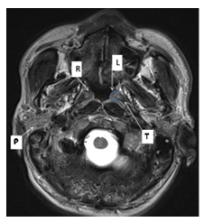
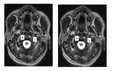
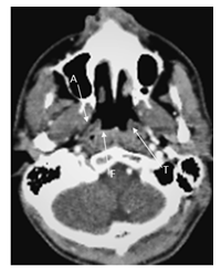
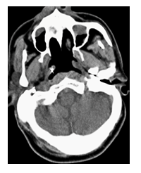
It may be debatable on the status of the newly described entity among researchers. But the structure appears anatomically coherent and can be identified on diagnostic imaging. A preliminary retrospective assessment by this author on a select number of cases of axial T2 weighted images including spin-echo images and FLAIR can identify the structure in the anatomical location which is isointense to the normal parotid gland. It can be otherwise described as slightly hyperintense to muscle, bone, and cartilage. Their location as per the original description is marked in Figure 1 and Figure 2. At the same time, routine non-contrast and contrast CT scans fail to specifically identify the structure described as tubarial gland (Figure 3 and Figure 4). This may be due to the limited inherent resolution of conventional CT images for such small structures. The same was true with noncontrast and postcontrast T1 weighted images. Even though the exact size in a normal individual may be difficult to define by imaging due to the submucosal location and the similar signal characters as the overlying mucosa, their presence appears consistent on MRI. It may be difficult to identify the structure in children due to the prominent nasopharyngeal wall mostly due to large adenoid tissue which has a similar signal character. It is seen to be non-conspicuous in elderly subjects, may be related to atrophy or poor contrast with adjacent structures.

Figure 1: Tubarial glands: Axial T2 weighted MR image of the base of the skull region and the nasopharynx showing the tubarial glands as per the description in the literature [1]. R and L indicate the right and the left tubarial glands which are posteromedial to torus tubaris (T). Approximate margin of the left gland is marked by a blue line. Note the signal characters of the right parotid gland (P).

Figure 2: Tubarial glands: Axial T2 weighted image at the nasopharynx and the palate level showing tapering gland on right (R)) and left (L) side. An outline of the gland is marked as per the literature description [1] on the left side mirror image.

Figure 3: CT image of nasopharynx: Post contrast axial CT image at the level of nasopharynx. T indicates the left torus tubaris, F indicates the right fossa of Rosenmüller and A indicates the right auditory tube.

Figure 4: Noncontrast CT image of the torus tubaris: Axial noncontrast CT image of 62 year old male showing a prominent nasopharyngeal lumen with clear demarcation of torus tubaris (white star).No demonstrable glandular elements could be noted around it.
Just like the recent discovery of the mesentery as the body’s 79th organ by J. Calvin Coffey [16] as recently in 2017 and subsequent endorsements including in anatomy textbooks like Gray’s Anatomy, this potential ‘organ 80’ may be endorsed later by the medical community [17]. Some researchers challenged the status of the newly described tubarial glands by citing the long history of known minor salivary glands in the nasopharynx. They contest this labeling of a macroscopic gland and maintain that these glandular elements merely indicate aggregation of many minor salivary glands in the area [18]. They maintain that an aggregate uptake of prostate-specific membrane antigen ligands in PET/CT scan alone may not be sufficient to attribute a major organ status. Some of the claims in the article of Valstar et al. were contested by some authors especially on overlooking several descriptions of the minor salivary glands around the auditory tube as early as 1837. They question the status of tubarial glands as the fourth pair of the major salivary gland but agree in designing radiotherapy fields to avoid these glands to preserve salivary function [19]. More research must be needed to settle these controversies and to formulate a consensus. Literature is scant in this field as the entity is just recently described. This author, however views that the description of this gland as tubarial gland is worthy and descriptive, whether as an aggregate of minor glands or as a major salivary gland. The pathologies of this entity may be discretely identified on imaging.
2.3 Minor salivary gland pathologies
Hundreds of scattered minor salivary glands (MSGs) in the head and neck region are a source of lubrication of their adjacent mucosa and immunity defense system; MSGs are also involved in a wide variety of pathologies. They are also an important source of diagnosing many diseases as many substances are actively secreted by the glands [5]. They are an important tissue source for diagnosing many oral and non-oral pathologies. Ease of excision of these glands and their negligible impact on the normal secretion capability of the salivary gland system are important benefits for such diagnostic tests. These glands are the site of several non-neoplastic pathologies including developmental, reactive, metabolic, inflammatory, immunologic diseases, iatrogenic causes, and conditions of unknown etiology [20]. Some of the common non-neoplastic conditions of MSGs are summarized in Table 1.
Demonstration of glandular amyloid deposits in the minor salivary gland by biopsy is valuable to confirm the diagnosis (sensitivity of 75%) of familial amyloidotic polyneuropathy [21]. Minor salivary gland biopsy (often from the labial area) is also valuable for the diagnosis of neonatal hemochromatosis [22], impaired lipid metabolism [23], scleroderma/systemic sclerosis [19], Sjogren syndrome [24], sarcoidosis [25] and IgG4-related disease [20, 26] in symptomatic patients. It is a less invasive procedure, safe, and easy to execute with minimal discomfort to the patient [22]. Van der Woude syndrome, Acute necrotizing sialometaplasia, Allergic sialadenitis, Orofacial granulomatosis, and Cheilitis glandularis are rare pathologies involving minor salivary glands [20]. Systemic diseases like graft versus host disease, IgG4-related disease, sarcoidosis, scleroderma/systemic sclerosis, and Sjogren syndrome may involve minor salivary glands which may cause xerostomia [20, 27].
Mucocele is a common lesion of the MSGs which present as a benign mucus-containing cyst directly under the mucosa. They can be a mucus extravasation cyst or a mucus retention cyst. Mucocele occurs due to rupture or blockage of a salivary gland duct. Common locations of mucocele are the lower lip, tongue, floor of the mouth (called ranula), and the buccal mucosa. Being a common benign pathology, they may need to be differentiated from more serious pathologies and mimics [28, 29]. Adenomatoid hyperplasia of the minor salivary gland is a rare non-neoplastic hyperplastic lesion frequently seen in the palate and rarely in the buccal mucosa. It is an asymptomatic tumor-like mass present as a mucosal swelling, often showing bluish color, and is due to localized hyperplasia of the minor salivary gland with normal glandular tissue on microscopy. The etiology of the entity is unknown and can clinically mimic salivary gland neoplasm [30].
Apart from the usual location of MSGs in the head and neck region which are scattered in the oral cavity, sinonasal cavities, oropharynx, larynx, and trachea; heterotopic minor salivary glands can also occur at many uncommon sites like lymph nodes, the capsule of the thyroid gland, facial bones or in the adenohypophysis. Any type of salivary gland tumors, either benign or malignant, can occur at any of these sites, including the heterotopic locations. Unlike the major salivary glands where approximately 80% of tumors are benign, malignant tumors are more common in minor salivary glands with reported incidences ranging from 40% to 80% [31-35]. Tumors of MSGs origin are uncommon, accounting for 23% of all malignant neoplasm of the upper aerodigestive tract and less than 20% of all salivary gland tumors [35].
The majority of MSGs are concentrated at the junction of the hard and soft palate. Thus, this region is the most common site for minor salivary gland tumors. 55% of tumors occur in the palate while the upper lip accounts for 15% of such tumors. The rest is distributed among the other glands in the floor of the mouth, retromolar region, cheek, tongue, peritonsillar area. Uncommonly, tumors may occur in the mandible apparently as intraosseous growth [33, 36].
Other benign tumors of minor salivary glands include Warthin's tumor, monomorphic adenomas, basal cell adenoma, and canalicular adenoma [37]. WHO recognized benign epithelial tumors of salivary glands [38] are summarized in Table 2. Pleomorphic adenomas carry a risk for malignant transformation (5-15%) and rarely with basal cell adenomas and Warthin tumors, while the other benign tumors in the list never develop into a malignancy. Pleomorphic adenoma has a rather high risk for recurrence which is extremely rare in other benign tumors of MSGs [31]. Swelling is the most common sign of MSGs tumors. Ulceration, ill-fitting dentures, difficulty of speaking, and pain are other presentations [39, 40].
|
Developmental pathologies |
Familial amyloidotic polyneuropathy Neonatal hemochromatosis Van der Woude syndrome |
|
Reactive lesions |
Acute necrotizing sialometaplasia Mucoceles |
|
Metabolic |
Altered lipid metabolism |
|
Inflammatory/ immunologic conditions |
Allergic sialadenitis Graft versus host disease IgG4-related disease Orofacial granulomatosis Scleroderma /systemic sclerosis Sjögren syndrome |
|
Iatrogenic |
Hyaluronic acid foreign body reaction |
|
Unknown/Undefined etiology |
Adenomatoid hyperplasia Cheilitis glandularis Sarcoidosis |
Table 1: Common non-neoplastic benign conditions affecting the minor salivary glands: Based on the probable etiology.
|
Pleomorphic adenoma myoepithelioma |
|
Basal cell adenoma (membranous, solid, trabecular and tubular types; often mixtures thereof) Warthin tumor |
|
Oncocytoma |
|
Lymphadenoma (sebaceous and non-sebaceous lymphadenoma) cystadenoma |
|
Sialadenoma papilliferum |
|
Ductal papilloma (intraductal and inverted ductal papilloma) Sebaceous adenoma |
|
Canalicular adenoma |
Table 2: Benign minor salivary gland tumors: WHO classification of the benign epithelial salivary gland tumors [32, 39].
In a large series (of 426 oral minor salivary gland tumors), Waldron et al. reported 57.5% benign and 42.5% malignant or potentially malignant tumors in their series. The mean age of all tumors was 50-60 year age group. The palate was the most common site followed by the upper lip and the buccal mucosa together accounted for 76.1% of all cases. The commonest benign tumor was pleomorphic adenoma (41% of all cases and 71% of all benign tumors) followed by monomorphic adenoma of the canalicular and basal cell subtypes (10% of all tumors and 18.9% of benign lesions) [37]. In another series, Wei-Yung et al. reported 119 minor salivary gland tumors, of which 56% were benign and 44% malignant. Pleomorphic adenoma was the commonest benign tumor (78%), followed by canalicular adenoma (21%). Mucoepidermoid carcinoma (45%), adenoid cystic carcinoma (23%), and low-grade adenocarcinoma (19%) were common malignancies [41].
Pleomorphic adenoma is the most common benign salivary
gland tumor (60%) of major and MSGs. The common site of MSGs involvement is the palate, followed by the upper lip and buccal mucosa. Other unusual sites are sinuses, larynx, epiglottis, trachea, tongue, soft palate, uvula, external auditory canal, etc. Clinically patient may present with a painless, slow-growing, well-circumscribed lump. Large tumors can cause mechanical symptoms depending on their location like dyspnoea, dysphagia, acute airway obstruction, and obstructive sleep apnea, epistaxis, nasal obstruction, etc [42-44]. Imaging with ultrasound, MRI, or computed tomography (CT) may be used depending on the site and size of the tumor [43]. They may appear as a well-defined, multilobulated mass on CT, and MRI. Calcification may be noted in CT images. On MRI, they are T2 hyperintense tumors with nodular enhancement and often with a T2 dark rim [43, 45]. Cytology and biopsy typically show a tumor of mixed epithelial cells and mesenchymal elements predominantly myoepithelial cells with no pseudocapsule. Differentiation from adenoid cystic carcinoma and polymorphous low-grade adenocarcinoma may be difficult with FNAC alone in some cases [44, 45].
Warthin tumors are commonly seen in the parotid tail region with occasional case reports of involvement of MSGs. They are associated with smoking and can be multiple. They are cystic, solid, or mixed slid-cystic tumors with very high metabolic activity on 18-fluorodeoxyglucose (18FDG)-PET and can mimic malignancy [45]. Approximately 80% of basal cell adenomas (BCAs) occur in major salivary glands and some cases in the oral cavity. Monomorphic adenomas are rare in MSGs. In a non-Asian population, canalicular adenoma is the most common adenoma of the benign tumors, with an incidence similar to myoepithelioma, 5-12%, of all benign tumors. Incidence varies widely with study location [31].
All salivary gland carcinomas constitute only 3-5% of head and neck malignancies. But they are the most diverse group of malignancy in the region with about 24 different WHO (World Health Organization) recognized types [46, 47] and are summarized in Table 3. The rarity combined with this enormous diversity of the tumor types and the in general unpredictability in the long-term outcome of these tumors poses a significant challenge in the management of salivary gland malignancies [48]. For formulating prognosis and treatment, unified clinical and pathologic parameters and meaningful therapeutic categorization are needed [49].
|
Low risk |
High risk |
|
Acinic cell carcinoma |
Sebaceous carcinoma and lymphadenocarcinoma |
|
Adenocarcinoma NOS and Cystadenocarcinoma, low grade |
Adenocarcinoma and cystadenocarcinoma, NOS, high grade |
|
Basal cell adenocarcinoma |
Small cell carcinoma |
|
Clear cell carcinoma |
Squamous cell carcinoma |
|
Carcinoma ex pleomorphic adenoma (intracapsular/minimally invasive or with low grade histology |
Carcinoma ex pleomorphic adenoma(widely invasive or high grade histology) |
|
Epithelial-myoepithelial carcinoma |
Adenoid cystic carcinoma |
|
Low grade salivary duct carcinoma (low grade cribriform cystadenocarcinoma) |
Large cell carcinoma |
|
Low grade mucoepidermoid carcinoma |
High grade mucoepidermoid carcinoma |
|
Oncocytic carcinoma |
Metastasizing pleomorphic adenoma |
|
Polymorphous low grade adenocarcinoma |
Mucinous adenocarcinoma |
|
Sialoblastoma |
Carcinosarcoma |
|
Myoepithelial carcinoma |
Lymphoepithelial carcinoma |
Table 3: Malignant minor salivary gland tumors: WHO recognized salivary gland malignancies based on the risk potential.
2.4 Potential tubarial gland pathologies
Both major and minor salivary glands produce similar pathologies including neoplasm. Hence, similar diseases may be expected in the case of tubarian glands. Most tumors arising in the nasopharynx are malignant with only rare benign nasopharyngeal tumors. Otitis media with effusion is very common with such malignant tumors but rare in benign tumors of the nasopharynx [50]. A retrospective analysis of a select few reported cases of MSGs origin tumors of the nasopharynx may give a glimpse of the pathology of this unsettled anatomical entity. More such research may be initiated once the status of the tubarian gland is settled.
Srinivas et al. reported a large nasopharyngeal benign minor salivary gland tumor characterized as pleomorphic adenoma. It was a 5.8 × 5.1 × 3.9 cm sized nasopharyngeal mass obliterating the fossa of Rosenmüller on the left side and causing severe narrowing of the nasopharynx. It was a solid enhancing mass with nodular calcifications. On retrospective analysis, this author believes that the tumor is located exactly in the expected location of the tubarial gland which extends from the base of skull level inferiorly [51]. A similar case of nasopharyngeal pleomorphic adenoma was reported by Garcia in a 61-year-old man, which was initially misdiagnosed as nasopharyngeal carcinoma. A review of the description and imaging features by this author can trace the epicenter of the mass as the right tubarial gland. This patient was evaluated by PET/CT which identified the right side nasopharyngeal tumor due to a larger and high intense area of uptake. Uptake of tracer was noted on the left side of the nasopharynx in proportion to the expected size of a normal tubarial gland, which was disregarded and reported as no uptake [52].
Jin et al. reported a small nasopharyngeal basal cell adenocaricinoma [53] in a 57-year-old man, which was the first of its kind reported in the area [53, 54]. It was a small infiltrating solid enhancing mass in the posterior nasopharynx anterior to torus tubaris measuring about 2.0 × 1.5 × 2.0 cm with a right retropharyngeal lymph node. The tumor extended across the right parapharyngeal space and infiltrated the medial pterygoid muscle and skull base. The case was managed by biopsy (which did not identify any salivary gland elements), followed by radiotherapy. On retrospective analysis, this author believes that the tumor is located exactly in the expected location of the tubarial gland which just expanded outside its normal boundary engulfing the gland. The tumor was originally attributed as of minor salivary gland origin. Basal cell adenocarcinoma (BCAC) of the minor salivary gland is an extremely rare malignancy with about 72 cases reported in the literature [53, 54]. Distant metastasis is rare (about 10%) with reports of liver metastasis [55]. Hwang et al. in 2020 reported a case of large, bilateral oncocytic cysts in the nasopharynx of minor salivary gland origin, which caused eustachian tube obstruction and chronic otitis media with effusion. It showed a bilateral lobulated submucosal cystic mass in front of torus tubaris obstructing the Eustachian tube opening [56]. Review of the available MRI images of the case by this author can be reinterpreted as enlarged bilateral tubarial glands affected by oncocytic cysts.
An extensive retrospective review of old cases of reported minor salivary was avoided for this unsettled entity. Establishing the status of the new gland in the description may broaden the understanding and characterize the lesions of the nasopharynx. Imaging changes of this gland with age, physiological changes, and post-radiation changes are also to be refined.
4. Conclusion
Identification of the nasopharyngeal salivary gland as a specific entity of the tubarial gland seems significant. This may be worthy to initiate more studies on the structure, importance, and diseases. This descriptive term may be a worthy addition to existing practice, till a consensus is reached on the status of the tubarian gland, either as a new organ, a new pair of major salivary gland, a part of the salivary gland system, a specific aggregate of nasopharyngeal minor salivary glands collectively labeled as tubarial glands or maintaining the status quo just as any other minor salivary gland. Understanding the role of the minor salivary gland seems important to preserve their function as that can be hampered in radiotherapy or surgery.
Conflict of Interest
Authors have no funding, financial or other conflicts of interest, including competing interests to disclose. This research did not receive any specific grant from funding agencies in the public, commercial or not-for-profit sectors.
References
- Valstar MH, de Bakker BS, Steenbakkers RJHM, et al. The tubarial salivary glands: A potential new organ at risk for radiotherapy. Radiother Oncol 20 (2020): 30809-4.
- Holsinger F, Bui D. Anatomy, function and evaluation of the salivary glands. In Eds.: Myers E, Ferris R. Salivary gland disorders. Springer-Verlag Berlin Heidelberg (2007): 1-16.
- Tuckers A, Ekstroem J, Khosravani N. Embryology and clinical anatomy; Regulatory mechanisms and salivary gland functions. In Eds.: Bradley P, Guntinas-Lichius O. Salivary gland disorders and diseases: diagnosis and management 1st Thieme, Stuttgart and New York (2011): 180.
- Tomoda K, Morii S, Yamashita T, et al. Deviation with increasing age in histologic appearance of submucosal glands in human eustachian tubes. Acta Otolaryngol 92 (1981): 463-467.
- Janice M Yoshizawa, Christopher A Schafer, Jason J Schafer, et al. Salivary Biomarkers: Toward Future Clinical and Diagnostic Utilities. Clinical Microbiology Reviews 26 (2013): 781-791.
- Tiwari M. Science behind human saliva. J. Nat. Sci. Biol. Med 2 (2011): 53-58.
- Seunghee Cha. Salivary Gland Development and Regeneration: Advances in Research and Clinical Approaches to Functional Restoration. Germany: Springer International Publishing (2017): 76.
- Hand AR, Pathmanathan D, Field RB. Morphological features of the minor salivary glands. Arch Oral Biol 44 (1999): 3-10.
- Eliasson L, Carlén A. An update on minor salivary gland secretions. Eur J Oral Sci 118 (2010): 435-442.
- Tos M. Mucous glands in the developing human rhinopharynx. Laryngoscope 87 (1977): 987-995.
- Frick H, Leonhardt H, Starck D. Human anatomy, 1st Thieme (1991).
- Klein Nulent, Valstar TJW, de Keize MH, et al. Physiologic distribution of PSMA-ligand in salivary glands and seromucous glands of the head and neck on PET/CT. Oral Surg Oral Med Oral Pathol Oral Radiol 125 (2018): 478-486.
- Bradley P, Guntinas-Lichius O. Salivary gland disorders and diseases: diagnosis and management, 1st, Thieme Publishing Group, Stuttgart and New York (2011).
- Radfar L, Sirois DA. Structural and functional injury in minipig salivary glands following fractionated exposure to 70 Gy of ionizing radiation: an animal model for human radiation-induced salivary gland injury. Oral Surg Oral Med Oral Pathol Oral Radiol Endod 96 (2003): 267-274.
- Demirci E, Sahin OE, Ocak M, et al. Normal distribution pattern and physiological variants of 68Ga-PSMA-11 PET/CT imaging. Nucl Med Commun 37 (2016): 1169-1179.
- Coffey JC, O’Leary DP. The mesentery: structure, function, and role in disease. Lancet Gastroenterol Hepatol 1 (2016): 238-247.
- Spring Hatfield RDH. Tubarial Salivary Glands: Scientists Discover New Salivary Glands in Human Throat. Todays RDH (2020).
- Guntinas-Lichius O, Ihrler S, Freesmeyer M, et al. Is there a new salivary gland? - Rather not! Laryngo- Rhino- Otologie 100 (2021): 12-14.
- lbert Mudry, Robert K Jackler. Are “Tubarial salivary glands” a previously unknown structure? Letter to the Editor. Radiotherapy and Oncology (2020).
- Doron J Aframian, Naama Keshet, Chen Nadler, et al. Minor salivary glands: Clinical, histological and immunohistochemical features of common and less common pathologies. Acta Histochemica 121 (2019): 151451.
- de Paula Eduardo F, de Mello Bezinelli L, de Carvalho DLC, et al. Minor salivary gland biopsy for the diagnosis of familial amyloid polyneuropathy. Neurol Sci 38 (2017): 311-318.
- Smith SR, Shneider BL, Magid M, et al. Minor salivary gland biopsy in neonatal hemochromatosis. Arch Otolaryngol Head Neck Surg 130 (2004): 760-763.
- Lukach L, Mali A, Zini Avraham, et al. Morphometrical study of minor salivary gland in xerostomic patients with altered lipid metabolism. Oral diseases 20 (2013): 10.
- Bamba R, Sweiss NJ, Langerman AJ, et al. The minor salivary gland biopsy as a diagnostic tool for Sjogren syndrome. Laryngoscope 119 (2009): 1922-1926.
- Harvey J, Catoggio L, Gallagher PJ, et al. Salivary gland biopsy in sarcoidosis. Sarcoidosis 6 (1989): 47-50.
- Doe K, Nozawa K, Okada T, et al. Usefulness of minor salivary gland biopsy in the diagnosis of IgG4-related disease: a case report. International journal of clinical and experimental pathology 7 (2014): 2673-2677.
- Matin M Imanguli, Jane C Atkinson, Sandra A Mitchell, et al. Salivary Gland Involvement in Chronic Graft-Versus-Host Disease: Prevalence, Clinical Significance, and Recommendations for Evaluation, Biology of Blood and Marrow Transplantation 16 (2010): 1362-1369.
- Senthilkumar B, Mahabob MN. Mucocele: An unusual presentation of the minor salivary gland lesion. Journal of pharmacy & bioallied sciences 4 (2012): 180-182.
- Lee KH, Cepeda L, Miller M, et al. Mucoceles not - Oral cysticercosis and minor salivary gland adenocarcinoma: Two case reports. Dermatology Online Journal 15 (2009): 8.
- Dereci Ömür, Cimen E. Adenomatoid hyperplasia of the minor salivary glands on the buccal mucosa: A rare case report. International journal of surgery case reports 5 (2014): 274-276.
- Hellquist H, Paiva-Correia A, Vander Poorten V, et al. Analysis of the Clinical Relevance of Histological Classification of Benign Epithelial Salivary Gland Tumours. Advances in therapy 36 (2019): 1950-1974.
- Eveson JW, Cawson RA. Tumors of the minor (oropharyngeal) salivary glands: a demographic study of 336 cases. J Oral Pathol 14 (1985): 500-509.
- Mishra S, Mishra YC. Minor salivary gland tumors in the Indian population: A series of cases over a ten year period. Journal of oral biology and craniofacial research 4 (2014): 174-180.
- Grazyna Wyszynska-Pawelec, Michal Gontarz, Jan Zapala, et al. Minor Salivary Gland Tumours of Upper Aerodigestive Tract: A Clinicopathological Study. Gastroenterology Research and Practice (2012): 780453.
- Rodney RM, Nicholas JC. Minor salivary gland tumors. In Eds.: Million RR, Caaassisi NJ. Management of Head and Neck Cancer: A Multidiscplinary Approach. 2nd J.B. Lippincott; Philadelphia (1994): 737.
- Lucas RB, Cawson RA, William H Binnie, et al. Lucas's Pathology of tumors of the oral tissues. Churchill Livingstone (1998).
- Waldron CA, El Mofty S, Gnepp DR. Tumors of the intraoral minor salivary glands: a demographic and histologic study of 426 cases. Oral Surg Oral Med Oral Pathol 66 (1988): 323-333.
- El-Naggar AK, Chan JKC, Grandis JR, et al. Tumours of salivary glands. In: WHO classification of head and neck tumours, 4th Lyon: IARC (2017): 159-202.
- Sarmento DJ, Morais ML, Costa AL, et al. Minor intraoral salivary gland tumors: a clinical-pathological study. Einstein (Sao Paulo, Brazil) 14 (2016): 508-512.
- Guzzo M, Locati LD, Prott FJ, et al. Major and minor salivary gland tumors. Crit Rev Oncol Hematol 74 (2010): 134-148.
- Wei-Yung Yih, James Kratochvil F, Jeffery CB. Stewart. Intraoral Minor Salivary Gland Neoplasms: Review of 213 Cases. Journal of Oral and Maxillofacial Surgery 63 (2005): 605-610.
- Grazyna Wyszynska-Pawelec, Michal Gontarz, Jan Zapala, et al. Minor Salivary Gland Tumours of Upper Aerodigestive Tract: A Clinicopathological Study. Gastroenterology Research and Practice 2012 (2012): 780453.
- Sahoo NK, Rangan MN, Gadad RD. Pleomorphic adenoma palate: Major tumor in a minor gland. Annals of maxillofacial surgery 3 (2013): 195-197.
- Kajiyama A, Edo H, Inoue N, et al. Magnetic Resonance Imaging and Histopathology in a Case of Pleomorphic Adenoma of a Minor Salivary Gland in the Nasal Cavity. The American journal of case reports 20 (2019): 679-684.
- Daniel Thomas Ginat. Imaging of Benign Neoplastic and Nonneoplastic Salivary Gland Tumors. Neuroimaging Clinics of North America 28 (2018): 159-169.
- Eveson JW, Auclair PL, Gnepp DR, et al. Tumors of the salivary glands: introduction. In Eds.: Barnes EL, Eveson JW, Reichart P, et al. World Health Organization classification of tumours: pathology & genetics. Head and neck tumours. Lyon: IARC Press (2005): 221-222.
- Seifert G, Brocheriou C, Cardesa A, et al. WHO International Histological Classification of Tumours. Tentative Histological Classification of Salivary Gland Tumours. Pathol Res Prac 186 (1990): 555-581.
- Bell RB, Dierks EJ, Homer L, et al. Management and outcome of patients with malignant salivary gland tumors. J Oral Maxillofac Surg 63 (2005): 917-928.
- Seethala RR. An Update on Grading of Salivary Gland Carcinomas. Head and Neck Pathol 3 (2009): 69-77.
- Maruyama A, Tsunoda A, Takahashi M, et al. Nasopharyngeal pleomorphic adenoma presenting as otitis media with effusion: case report and literature review. American Journal of Otolaryngology 35 (2014): 73-76.
- Srinivas CV, Mahesh A, Nair S. Nasopharyngeal Pleomorphic Adenoma: A Difficult Surgical Proposition 11 (2017): MD01-MD02.
- Garcia GJA. Misdiagnosis of a Nasopharyngeal Pleomorphic Adenoma as a Nasopharyngeal Carcinoma. J Otolaryngol ENT Res 1 (2014).
- Jin J, He XY. Basal cell adenocarcinoma of the nasopharyngeal minor salivary glands: a case report and review of the literature. BMC cancer 18 (2018): 878.
- Cuthbertson DW, Raol N, Hicks J, et al. Minor salivary gland basal cell adenocarcinoma: a systematic review and report of a new case. JAMA Otolaryngol Head Neck Surg 141 (2015): 276-283.
- Chen S, Yang S, Chen X. Basal cell adenocarcinoma of the buccal minor salivary gland with liver metastases. Ann Saudi Med 35 (2015): 318-320.
- Joshua C Hwang, Raj D Dedhia, Joan E Bernard, et al. Oncocytic Cysts of the Nasopharynx: A Case Report Allergy & Rhinology 11 (2020).
