Impact of Acceleration on Human Visual Acuity and Stereopsis
Article Information
Sunina A1, Ramachandra V Shet2 *, Rajib Mandal3, Yogesh Vaghela4
1Intern B.Sc Optometry student, A.J Institute of Allied Health Sciences, Kuntikana, Mangalore
2HOD, Department of Optometry, A.J Institute of Allied Health Sciences, Kuntikana, Mangalore
3Principal, Netra Jyothi Institute of Allied Health Sciences, Udupi, Karnataka, India
4Assistant professor and Research Fellow, Charotar University of Science and Technology
*Corresponding Author: Ramachandra V Shet, HOD, Department of Optometry, A.J Institute of Allied Health Sciences, Kuntikana, Mangalore
Received: 21 May 2021; Accepted: 27 May 2021; Published: 09 June 2021
Citation: Sunina A, Ramachandra V, Rajib Mandal, Yogesh Vaghela. Impact of Acceleration on Human Visual Acuity and Stereopsis. Journal of Environmental Science and Public Health 5 (2021): 331-341.
Share at FacebookAbstract
Purpose: To assess the impact of horizontal acceleration on human visual acuity and stereopsis.
Methods: The written consent of 60 subjects (36 females and 24 males) aged between 18-24 years was taken after proper explanations. History taking followed by VA measurement, refraction, slit lamp examination, Ophthalmoscopy was performed to rule out the exclusion criteria. Measurements of near visual acuity and stereopsis was done in 3 speeds, slow, moderate and high speed of tread mill for all subjects. Static and dynamic VA measurements was noted. Dynamic VA was measured in tread mill with examiner holding the VA chart. After completing the near visual acuity examination, participant was subjected to stereopsis test using the stereo-test circles with both eyes. Duration of each test was noted.
Results: The ANNOVA test analysis revealed no significant difference in visual acuity and stereopsis measured in slow and moderate speed in comparison with static visual acuity and stereopsis (p>0.05). However, a highly significant difference was found in visual acuity and stereopsis measured during high speed (p<0.01). Also, it was observed that time taken to read visual acuity and stereopsis chart increased with increasing speed (p<0.01). It was also found that majority of subjects experienced different level of dizziness, headache, ocular discomfort during acceleration.
Conclusion: High acceleration had a highly significant effect on visual acuity and stereopsis.
Keywords
Visual acuity; Stereo acuity; Dynamic; Static; Tread mill; Visual discomfort; Headache; Acceleration
Visual acuity articles; Stereo acuity articles; Dynamic articles; Static articles; Tread mill articles; Visual discomfort articles; Headache articles; Acceleration articles
Visual acuity articles Visual acuity Research articles Visual acuity review articles Visual acuity PubMed articles Visual acuity PubMed Central articles Visual acuity 2023 articles Visual acuity 2024 articles Visual acuity Scopus articles Visual acuity impact factor journals Visual acuity Scopus journals Visual acuity PubMed journals Visual acuity medical journals Visual acuity free journals Visual acuity best journals Visual acuity top journals Visual acuity free medical journals Visual acuity famous journals Visual acuity Google Scholar indexed journals Stereo acuity articles Stereo acuity Research articles Stereo acuity review articles Stereo acuity PubMed articles Stereo acuity PubMed Central articles Stereo acuity 2023 articles Stereo acuity 2024 articles Stereo acuity Scopus articles Stereo acuity impact factor journals Stereo acuity Scopus journals Stereo acuity PubMed journals Stereo acuity medical journals Stereo acuity free journals Stereo acuity best journals Stereo acuity top journals Stereo acuity free medical journals Stereo acuity famous journals Stereo acuity Google Scholar indexed journals Dynamic articles Dynamic Research articles Dynamic review articles Dynamic PubMed articles Dynamic PubMed Central articles Dynamic 2023 articles Dynamic 2024 articles Dynamic Scopus articles Dynamic impact factor journals Dynamic Scopus journals Dynamic PubMed journals Dynamic medical journals Dynamic free journals Dynamic best journals Dynamic top journals Dynamic free medical journals Dynamic famous journals Dynamic Google Scholar indexed journals Static articles Static Research articles Static review articles Static PubMed articles Static PubMed Central articles Static 2023 articles Static 2024 articles Static Scopus articles Static impact factor journals Static Scopus journals Static PubMed journals Static medical journals Static free journals Static best journals Static top journals Static free medical journals Static famous journals Static Google Scholar indexed journals Tread mill articles Tread mill Research articles Tread mill review articles Tread mill PubMed articles Tread mill PubMed Central articles Tread mill 2023 articles Tread mill 2024 articles Tread mill Scopus articles Tread mill impact factor journals Tread mill Scopus journals Tread mill PubMed journals Tread mill medical journals Tread mill free journals Tread mill best journals Tread mill top journals Tread mill free medical journals Tread mill famous journals Tread mill Google Scholar indexed journals Visual discomfort articles Visual discomfort Research articles Visual discomfort review articles Visual discomfort PubMed articles Visual discomfort PubMed Central articles Visual discomfort 2023 articles Visual discomfort 2024 articles Visual discomfort Scopus articles Visual discomfort impact factor journals Visual discomfort Scopus journals Visual discomfort PubMed journals Visual discomfort medical journals Visual discomfort free journals Visual discomfort best journals Visual discomfort top journals Visual discomfort free medical journals Visual discomfort famous journals Visual discomfort Google Scholar indexed journals Headache articles Headache Research articles Headache review articles Headache PubMed articles Headache PubMed Central articles Headache 2023 articles Headache 2024 articles Headache Scopus articles Headache impact factor journals Headache Scopus journals Headache PubMed journals Headache medical journals Headache free journals Headache best journals Headache top journals Headache free medical journals Headache famous journals Headache Google Scholar indexed journals Acceleration articles Acceleration Research articles Acceleration review articles Acceleration PubMed articles Acceleration PubMed Central articles Acceleration 2023 articles Acceleration 2024 articles Acceleration Scopus articles Acceleration impact factor journals Acceleration Scopus journals Acceleration PubMed journals Acceleration medical journals Acceleration free journals Acceleration best journals Acceleration top journals Acceleration free medical journals Acceleration famous journals Acceleration Google Scholar indexed journals human visual articles human visual Research articles human visual review articles human visual PubMed articles human visual PubMed Central articles human visual 2023 articles human visual 2024 articles human visual Scopus articles human visual impact factor journals human visual Scopus journals human visual PubMed journals human visual medical journals human visual free journals human visual best journals human visual top journals human visual free medical journals human visual famous journals human visual Google Scholar indexed journals
Article Details
1. Introduction
Stereopsis is the capacity to see profundity of field dependent on the difference between the pictures framed by the two eyes-it is the most exceptional visual capacity. Wheatstone imagined the stereoscope and found that, if territories on the retina of the two eyes that are isolated on a level plane were animated at the same time, stereopsis could happen [1]. Visual sharpness is the most usually considered vision-related boundary that influences stereopsis. Stereopsis deteriorates in any event, when visual disability is available in just one eye [2-6].
Object recognition critically depends on motion perception which is associated with velocity and acceleration of target. Various acceleration mechanisms are seen in daily activities, such as walking, running, driving, travelling and working with some vehicles (cars, high-speed rails, rapid transit systems) which may create problems such as motion sickness and spatial disorientation. At exposure acceleration, a minimum amount of blood would return to heart and rest accumulate in lower limb, which result in hypoxia and ischemia of the brain and eye affect pilots with a group of visual impairment like; black-out, gray out, peripheral vision loss, unconsciousness after coming across into high acceleration. Objects appear as blur throughout rapid acceleration. Currently the effect of horizontal acceleration on visual acuity and stereopsis remains ambiguous. The exact vision changes from the acceleration of have been rarely studied. Chi- Ting Horng et al. while studying on 20 participants (mean age= 22.6 years) to understand horizontal acceleration and its impact upon human stereopsis and visual acuity and stereopsis; they have used acceleration from two different direction with two types of acceleration. i.e moderate higher than 0.1g and lower than 0.1g. Speed increases were created on a speeding up stage where the subjects stood. The visual acuity and stereopsis of the eye were measured before and during the acceleration. It was found that acceleration less than 0.1g did not affect dynamic vision and stereopsis. And also, vision decreased and stereopsis declined significantly when acceleration more than 0.1g [7]. Ming-Ling Tsai et al. evaluated the ocular responses and visual performance after high acceleration force exposure.14 men were enrolled in the study. A human centrifuge was used to induce 9 times the acceleration force in head to toe direction. Visual performance was evaluated using ETDRS visual chart and contrast sensitivity was examined before and after centrifugation. High acceleration force may induce transient visual acuity reduction and temporal corneal thickening. Prolonged increase in anterior chamber depth and pupillary dilation were also observed [8]. Joseph L. Demer et al. in their study about the effect of passive vertical head motion on dynamic visual acuity in 13 subjects they found that acuity declined with increasing velocity. Static visual acuity was measured without motion.
Dynamic visual acuity was measured through vertical sinusoidal motion of either optotypes or subject seating on a servo-driven rotating chair. Dynamic visual acuity for head motion was measured unaided, as well as with telescopic spectacles [9]. This study aims to understand the impact of horizontal acceleration on human visual acuity and stereopsis.
2. Methodology
In this cross-sectional study 60 emmetropic subjects (36 females and 24 males) aged between 18-24 years included in this study. Subjects with a history of ocular surgery, ocular diseases or systemic diseases were excluded from this study.
2.1 Methods
The study was started after obtaining ethical clearance from the Institutional Ethical Committee. In the first step demographic data of subjects were taken along with an informed consent form to fill. All subjects were gotten some information about the clinical history, any past visual infections or medical procedures and utilization of meds. Then they had undergone general examination like distant visual acuity testing using Snellen chart at 6m. Objectives as well as Subjective refraction were done to find out if the patients have any kind of refractive error. External examination like cover test Slit-lamp examination and fundus examination were performed to find out if there is any ocular disease present.
Visual acuity was measured using the standardized computer chart named Oscar vision chart. Distance visual acuity was measured using Snellen chart at a distance of 6m from patient to the monitor. The near visual acuity was also measured using the reduced Snellen chart at 33cm. Retinoscopy and subjective refraction were performed by maintaining standard protocol.
External examination was included the assessment of facial symmetry, behavioural and morphological characters and its association with any kind of systemic or ocular diseases. It also includes cover test, and which is performed at distance and near to evaluate tropia and phoria. Orthophoria was considered if there is no deviation observed. EXO indicate the outward deviation and ESO indicate the inward deviation. Slit lamp examination was done using the Zeiss type illumination system. The fundus examinations were done by using ophthalmoscope.
Near vision card was held at their normal working distance. Measurements of near visual acuity was done in 3 speeds, slow, moderate and high speed of tread mill. Two types of VA measurements were taken, dynamic and static. Static method of VA measurements was performed without involvement of treadmill. Dynamic VA measurements was taken in tread mill with patient holding the VA chart. The measurement of visual acuity was taken while the patient was reading the VA chart on the moving platform of tread mill. Time taken was noted down for each speed. Static and dynamic stereo acuity test was performed by using Circle test. Measurement was done in 3 speeds. Duration of each test was noted. Questionnaire was given after assessment of dynamic visual acuity at slow moderate and high speeds.
Statistical analysis was done by using ANOVA with Turkey HSD test. Chi- square test was performed to analyses differences of different parameter value as required, P value for confidence interval of 95% was considered significant at the P < 0.05 level for prevalence estimate.
3. Result
In present study out of 60 subjects in this study 36 were females and 24 were males. Mean age of subject was 20.933 + 1.24. The ANNOVA test analysis of the data revealed no significant difference in visual acuity and stereopsis measured in slow and moderate speed in comparison with static visual acuity and stereopsis (p>0.05). However, a highly significant difference was found in visual acuity stereopsis measured during high speed in comparison with static visual acuity and stereopsis (p<0.01). Also, it was observed that time taken to read visual acuity and stereopsis chart increased with increasing speed (p<0.01).

Table 1: Age distribution of subject
Graph 1: Age distribution of subject
Table 1 and Graph 1 shows that out of 60 individuals considered for this study majority of the subject 31 (51.7%) were of 21-22, followed by 23 (38.3%) subject were of 19-20 years and 6 (10.0%) were of 23-25 years.
Table 2 and Graph 2 shows that out of 60 individuals 36 (60%) subjects were female and 24 (40%) subjects were males.

Table 2: Gender distribution of subject
Graph 2: Gender distribution of subjects

Table 3: Religion distribution of subjects
Graph 3: Religion distribution of subjects

Table 4: Represents the mean value of the time taken for stereoacuity measurement
Graph 4: Represents the mean value of the time taken for stereoacuity measurement
Table 3 and Graph 3 shows that, out of 60 individuals considered for this study 29 (48%) were Hindus, 20 (34%) were Christians and 11 (18%) subjects were Muslims.
Table 4 and Graph 4 shows that the mean value of time taken for static stereoacuity measurement is 18.63 + 5.38 and mean value of time taken for stereoacuity measurement in slow speed is 21.78 + 5.96, moderate speed is 22.63 + 5.88, high speed is 23.24 + 5.88.
|
Parameter |
50 Arc |
60 Arc |
80 Arc |
100 Arc |
140 Arc |
200 Arc |
400 Arc |
Total |
P |
|
|
Static |
Count |
1 |
1 |
1 |
6 |
49 |
2 |
0 |
60 |
0.00 HS |
|
% |
1.7% |
1.7% |
1.7% |
10.0% |
81.7% |
3.3% |
0% |
100% |
||
|
Slow |
Count |
1 |
1 |
1 |
6 |
49 |
2 |
0 |
60 |
0.00 HS |
|
% |
1.7% |
1.7% |
1.7% |
10.0% |
81.7% |
3.3% |
0% |
100% |
||
|
Moderate |
Count |
1 |
1 |
1 |
6 |
49 |
2 |
0 |
60 |
0.00 HS |
|
% |
1.7% |
1.7% |
1.7% |
10.0% |
81.7% |
3.3% |
0% |
100% |
||
|
High |
Count |
0 |
0 |
2 |
1 |
7 |
48 |
2 |
60 |
0.00 HS |
|
% |
0.0% |
0.0% |
3.3% |
1.7% |
11.7% |
80.0% |
3.3% |
100% |
||
Table 5: Represents the frequency distribution of stereoacuity value.
Table 5 shows the distribution of stereoacuity value at different speeds. Majority of subjects (81.7%) had stereoacuity of 140 sec of arc at static, slow and moderate speed. In high speed the stereoacuity was reduced to 200 sec of arc in majority of subjects (80%).
Table 6: Represents the frequency distribution value of visual acuity.
Table 6 shows the distribution of visual acuity value at different speeds. Out of 60 subjects majority of subjects (100%) had visual acuity of N6 at static, slow speed. 57 subjects (95%) had a visual acuity of N6 and 3 subjects (5%) had a visual acuity of N8 at moderate speed. In high speed the visual acuity was reduced to N8 in 59 subjects (98.3%) and N12 in 1 subject (1.7%).

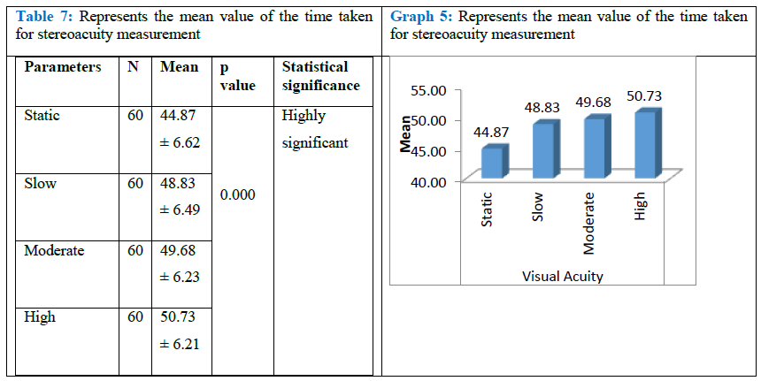
Table 7: Represents the mean value of the time taken for stereoacuity measurement
Graph 5: Represents the mean value of the time taken for stereoacuity measurement
Table 7 and Graph 5 shows that the mean value of time taken for static visual acuity measurement is 44.87 + 6.62 and mean value of time taken for visual acuity measurement in slow speed is 48.83 + 6.49, moderate speed is 49.68 + 6.23, high speed is 50.73 + 6.21.
|
1 |
2 |
|||
|
Count |
% |
Count |
% |
|
|
Q-1 |
0 |
.0% |
60 |
100.0% |
|
Q-2 |
24 |
40.0% |
36 |
60.0% |
|
Q-3 |
48 |
80.0% |
12 |
20.0% |
|
Q-4 |
28 |
46.7% |
32 |
53.3% |
|
Q-5 |
36 |
60.0% |
24 |
40.0% |
|
Q-6 |
0 |
.0% |
60 |
100.0% |
|
Q-7 |
58 |
96.7% |
2 |
3.3% |
|
Q-8 |
51 |
85.0% |
9 |
15.0% |
|
Q-9 |
0 |
.0% |
60 |
100.0% |
|
Q-10 |
60 |
100.0% |
0 |
.0% |
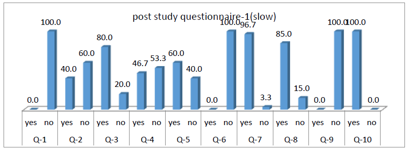
Table 8: Represents post study questionnaire 1.
Table-8 shows that, the Q-1 (100%-no) which states that all the subjects were not able to read , Q-2 showed pain in eyes while reading among 24 subjects (40%-yes) and 36 subjects ( 60%-no) , Q-3 showed tiredness in eyes while reading among 48 subjects (80%-yes) and 12 subjects (20%-no), Q-4 showed headache while reading among 28 subjects (46.7%-yes) and 32 subjects (53.3%-no), Q-5 showed giddiness while reading among 36 subjects (60%-yes) and 24 subjects (40%-no), Q-6 showed (100%-no) which states that here was no vomiting sensation while reading, Q-7 showed blurry vision while reading among 58 subjects (96.7%-yes) and 2 subjects (3.3%-no) , Q-8 showed difficulty moving eyes while reading among 51 subjects (85%-yes) and 9 subjects (15%-no) , Q-9 showed (100%-no) which states that here was no comfort response while reading, Q-10 showed (60%-yes) which states that there was heaviness in eyes after reading.

Graph 6: Represents post study questionnaire 1.
Graph-8 shows that, Q-1 (100%-no), Q-2 showed pain in eyes while reading among 24 subjects (40%-yes) and 36 subjects ( 60%-no), Q-3 showed 48 subjects (80%-yes) and 12 subjects (20%-no), Q-4 (46.7%-yes) and 32 subjects (53.3%-no), Q-5 showed 36 subjects (60%-yes) and 24 subjects (40%-no), Q-6 showed (100%-no), Q-7 showed 58 subjects (96.7%-yes) and 2 subjects (3.3%-no), Q-8 showed among 51 subjects (85%-yes) and 9 subjects (15%-no), Q-9 showed (100%-no), Q-10 showed (60%-yes).
|
2 |
4 |
Total |
||||
|
Count |
% |
Count |
% |
Count |
% |
|
|
Q-1 |
0 |
.0% |
60 |
100.0% |
60 |
100.0% |
|
Q-2 |
24 |
40.0% |
36 |
60.0% |
60 |
100.0% |
|
Q-3 |
48 |
80.0% |
12 |
20.0% |
60 |
100.0% |
|
Q-4 |
29 |
48.3% |
31 |
51.7% |
60 |
100.0% |
|
Q-5 |
36 |
60.0% |
24 |
40.0% |
60 |
100.0% |
|
Q-6 |
1 |
1.7% |
59 |
98.3% |
60 |
100.0% |
|
Q-7 |
58 |
96.7% |
2 |
3.3% |
60 |
100.0% |
|
Q-8 |
50 |
83.3% |
10 |
16.7% |
60 |
100.0% |
|
Q-9 |
0 |
.0% |
60 |
100.0% |
60 |
100.0% |
|
Q-10 |
60 |
100.0% |
0 |
.0% |
60 |
100.0% |
Table 9: Represents post study questionnaire 2.
|
2 |
4 |
Total |
||||
|
Count |
% |
Count |
% |
Count |
% |
|
|
Q-1 |
0 |
.0% |
60 |
100.0% |
60 |
100.0% |
|
Q-2 |
24 |
40.0% |
36 |
60.0% |
60 |
100.0% |
|
Q-3 |
48 |
80.0% |
12 |
20.0% |
60 |
100.0% |
|
Q-4 |
29 |
48.3% |
31 |
51.7% |
60 |
100.0% |
|
Q-5 |
36 |
60.0% |
24 |
40.0% |
60 |
100.0% |
|
Q-6 |
1 |
1.7% |
59 |
98.3% |
60 |
100.0% |
|
Q-7 |
58 |
96.7% |
2 |
3.3% |
60 |
100.0% |
|
Q-8 |
50 |
83.3% |
10 |
16.7% |
60 |
100.0% |
|
Q-9 |
0 |
.0% |
60 |
100.0% |
60 |
100.0% |
|
Q-10 |
60 |
100.0% |
0 |
.0% |
60 |
100.0% |
Table 10: Represents post study questionnaire 3.
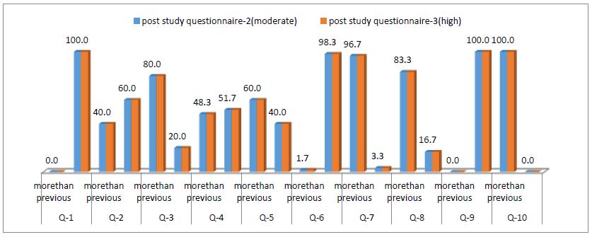
Table-8 and 9 shows that, Q-1 (100%-no) which states that all the subjects were not able to read in post study questionnaire 2 and 3, Q-2 showed pain in eyes while reading among 24 subjects (40%-more than previous) and 36 subjects (60%-No) in post study questionnaire 2 and 3, Q-3 showed tiredness in eyes while reading among 48 subjects (80%- more than previous) and 12 subjects (20%-no) in post study questionnaire 2 and 3, Q-4 showed headache while reading among 29 subjects (48.3%- more than previous) and 31 subjects (51.7%-no) in post study questionnaire 2 and 3, Q-5 showed giddiness while reading among 36 subjects (60%- more than previous) and 24 subjects (40%-no) in post study questionnaire 2 and 3, Q-6 showed 1 subject (1.7%- more than previous ) and 59 subjects (98.3%-no) which states that here was no vomiting sensation while reading in post study questionnaire 2 and 3, Q-7 showed blurry vision while reading among 58 subjects (96.7%- more than previous) and 2 subjects (3.3%-no) in post study questionnaire 2 and 3, Q-8 showed difficulty moving eyes while reading among 50 subjects (83.3%- more than previous) and 10 subjects (16.7%-no) in post study questionnaire 2 and 3, Q-9 showed (100%-no) which states that here was no comfort response while reading in post study questionnaire 2 and 3, Q-10 showed (60%- more than previous) which states that there was heaviness in eyes after reading in post study questionnaire 2 and 3.

Graph 9: Represents post study questionnaire 2 and 3.
Graph 9 showed no similar or less than previous.
All the subjects reported either no or more than previous response in moderate and high speed, it reported more than previous in all the subjects.
4. Discussion
Object recognition critically depends on motion perception which is associated with velocity and acceleration of target. Various acceleration mechanisms are seen in daily activities, such as walking, running, driving, travelling and working with some vehicles (cars, high-speed rails, rapid transit systems) which may create problems such as motion sickness and spatial disorientation.
Ching-Thing Horng found that acceleration less than 0.1g did not affect dynamic vision and stereopsis. But vision decreased and stereopsis declined significantly when acceleration more than 0.1g and also all subjects complained of discomfort and ocular strain while reading the letter cards and stereopsis picture. In present study I found that slow and moderate speed did not affect dynamic vision and stereopsis. But vision and stereopsis decreased in high speed and subjects experienced different level of discomfort and ocular strain. This study concluded that object cannot be distinctly differentiated if there is rapid head movement. Similarly, present study concluded head movements degrade the acuity by producing motion, particularly acceleration of the retinal images of stationary objects because of compensatory mechanisms. The relative position of human body and head may influence dynamic VA and stereopsis when humans are walking faster [7]. Ming-Ling Tsai et al. found High-speed increase power may incite transient visual keenness decrease and impermanent corneal thickening. Drawn out expansion in ACD and pupillary enlargement were additionally noticed. The decline in CS endured for 30 minutes after centrifugation. The systems hidden these perceptions are not satisfactory, on the grounds that there are no past writes about this point [8]. Current study also supports this statement. This study found a highly significant difference was found in visual acuity and stereopsis measured during high speed (p<0.01). Also, it was observed that time taken to read visual acuity and stereopsis chart increased with increasing speed (p<0.01). But current study didn’t take the measurement of ACD, Pupillary enlargement and CS. Current study is unable to comment on this point. It was also found that majority of subjects experienced different level of dizziness, headache, ocular discomfort during acceleration. In another study Joseph L. had also found out that during optotype motion acuity declined with increasing velocity which support result of current study [9].
Horng CT et al had done study on changes in visual function during the coriolis illusion. Coriolis illusion produces spatial orientation. The study included 18 subjects of mean age 24.7 years of age. A spatial confusion test system was utilized to deliver coriolis incitement. The visual sharpness of the subjects was assessed with the rosenbaum vision card previously and during coriolis incitement. Stereopsis was estimated with the titmus stereo-acuity test. Throughout the experiment, eyeball movements were observed on a television monitor. Electrooculography (EOG) and electroencephalography (EEG) were also document. They found that before coriolis stimulation, the visual acuity and stereopsis of all subjects were 20/20 and 40 sec of arc respectively. During the coriolis illusion visual acuity dropped by 2 lines. Stereopsis of subjects were also decreased. They concluded that head movements may affect visual acuity and stereopsis because of the incorrected retinal image that reveals poor capacity for macular sensory fusion [10]. Current study also concluded that time taken to read visual acuity and stereopsis chart increased with increasing speed.
Only a specific age group age group (18-25) were included in the study. This study was limited to small area of Mangalore with small sample size. As the subjects moves continuously on treadmill there is a chance of variation in working distance. These were few limitations in our study.
5. Conclusion
Present study concluded that high acceleration had a highly significant effect on visual acuity and stereopsis. But visual acuity and stereopsis was not affected by slow and moderate speed. People may go through different level of giddiness, headache, ocular irritations while concentrating to a particular object with acceleration.
References
- Wheatstone C. Contributions to the physiology of vision. Part the first. On some remarkable and hitherto unobserved phenomena of binocular vision. Philos. Trans. R. Soc. Lond. B Biol. Sci 371 (1838): 128.
- Lam AK, Chau AS, Lam WY, et al. Effect of naturally occurring visual acuity differences between two eyes in stereoacuity. Ophthalmic Physiol. Opt 16 (1996): 189-195.
- Donzis PB, Rappazzo JA, Burde RM, et al. Effect of binocular variations of Snellen’s visual acuity on Titmus stereoacuity. Arch. Ophthalmol 101 (1983): 930-932.
- Larson WL, Bolduc M. Effect of induced blur on visual acuity and stereoacuity. Optom. Vis. Sci 68 (1991): 294-298.
- Levy NS, Glick EB. Stereoscopic perception and Snellen visual acuity. Am. J. Ophthalmol 78 (1974): 722-724.
- Goodwin RT, Romano PE. Stereoacuity degradation by experimental and real monocular and binocular amblyopia. Invest. Ophthalmol. Vis. Sci 26 (1985): 917-923.
- Chi-Ting Horng, Yih-Shou Hsieh, Ming-Ling Tsai, et al. Effects of horizontal acceleration on human visual acuity and stereopsis. Int J Environ Res Public Health 12 (2015): 910-926.
- Ming-Ling Tsai, Chun-Cheng Liu, Yi-Chang Wu, et al. Ocular responses and visual performance after high-acceleration force exposure. Invest Ophthalmol Vis Sci 50 (2009): 4836-4839.
- Joseph L Demer, Firooz Amjadi. Dynamic visual acuity of normal subjects during vertical optotype and head motion.Invest Ophthalmol Vis Sci 34(1993): 1894-1906.
- Chi-Ting Horng, Chung-Cheng Liu, Daih-Iluang Kuo, et al. Changes in visual function during the Coriolis illusion. Aviat Space Environ Med 80(2009): 360-363.
