Imaging Cerebral Haemorrhage using MRI: Improved Sensitivity of Susceptibility Weighted Imaging (SWI) Compared to Gradient Echo Sequences (GRE)
Article Information
Punitha P2, Karunanithi Rajamanickam1*, Krishnamoorthy A2, Einstein A2, Alex Daniel Prabhu K2, Murugesan R1
1Faculty of Allied Health Sciences, Chettinad Academy of Research and Education, Kelambakkam, Tamilnadu, India
2Chettinad Hospital and Research Institute, Kelambakkam, Tamilnadu, India
*Corresponding author: Karunanithi Rajamanickam, Assistant Professor [Physics], Chettinad Academy of Research and Education, Rajiv Gandhi Salai (OMR), Kelambakkam, Chengalpattu District, Tamilnadu, 603103, India
Received: 24 December 2019; Accepted: 02 January 2020; Published: 08 January 2020
Citation:
Punitha P, Karunanithi Rajamanickam, Krishnamoorthy A, Einstein A, Alex Daniel Prabhu K, Murugesan R. Imaging Cerebral Haemorrhage using MRI: Improved Sensitivity of Susceptibility Weighted Imaging (SWI) Compared to Gradient Echo Sequences (GRE). Archives of Internal Medicine Research 3 (2020): 018-025.
Share at FacebookAbstract
Background: The appearance of cerebral haemorrhage (CH) as common MR imaging findings raise the question of how MR image acquisition and haemorrhage area quantification can help in understanding brain pathological changes as the result of various disease conditions such as acute brain injury, hypertension amyloid angiopathy and other diverse pathology. Neuroimaging is essential for the clinician to identify the cause of haemorrhage. In this study, we examined 25 cerebral haemorrhage MR images with an objective of comparing two different sequences viz., susceptibility weighted imaging (SWI) and gradient recalled echo (GRE) on same patients and also to quantify the area of haemorrhage using manual selection and gray area threshold (automated selection) in the digital image.
Materials and Methods: In this prospective study, MR images were acquired from 25 subjects with acute brain trauma (16) , hypertension (5), and amyloid angiopathy (4), using SWI and GRE MR Sequences, with section thicknesses (3- 5 mm), and magnetic field strengths (1.5T). Individual CH was manually identified and analyzed for haemorrhage area using signal intensity changes by manual and automatic selection of binary threshold hypointense signal by ImageJ software package.
Results: By other parameters set as constant, we were able to quantify hypointense signal changes at regions of cerebral haemorrhage on SWI when compared to no such notable changes in GRE in the case of amyloid angiopathy patients. Apart from this, lesions prospectively identified on SWI had a significantly greater area measured on the SWI image than those not prospectively identified on GRE.
Conclusions: There is an increased sensitivity in the CH detection in patients with amyloid angiopathy while using SWI sequence when compared to GRE in which we could not identify any signal changes in specific areas. Furt
Keywords
MR Sequences, SWI, GRE, Cerebral Haemorrhage, Binary Image, Image Threshold
MR Sequences articles MR Sequences Research articles MR Sequences review articles MR Sequences PubMed articles MR Sequences PubMed Central articles MR Sequences 2023 articles MR Sequences 2024 articles MR Sequences Scopus articles MR Sequences impact factor journals MR Sequences Scopus journals MR Sequences PubMed journals MR Sequences medical journals MR Sequences free journals MR Sequences best journals MR Sequences top journals MR Sequences free medical journals MR Sequences famous journals MR Sequences Google Scholar indexed journals SWI articles SWI Research articles SWI review articles SWI PubMed articles SWI PubMed Central articles SWI 2023 articles SWI 2024 articles SWI Scopus articles SWI impact factor journals SWI Scopus journals SWI PubMed journals SWI medical journals SWI free journals SWI best journals SWI top journals SWI free medical journals SWI famous journals SWI Google Scholar indexed journals GRE articles GRE Research articles GRE review articles GRE PubMed articles GRE PubMed Central articles GRE 2023 articles GRE 2024 articles GRE Scopus articles GRE impact factor journals GRE Scopus journals GRE PubMed journals GRE medical journals GRE free journals GRE best journals GRE top journals GRE free medical journals GRE famous journals GRE Google Scholar indexed journals Cerebral Haemorrhage articles Cerebral Haemorrhage Research articles Cerebral Haemorrhage review articles Cerebral Haemorrhage PubMed articles Cerebral Haemorrhage PubMed Central articles Cerebral Haemorrhage 2023 articles Cerebral Haemorrhage 2024 articles Cerebral Haemorrhage Scopus articles Cerebral Haemorrhage impact factor journals Cerebral Haemorrhage Scopus journals Cerebral Haemorrhage PubMed journals Cerebral Haemorrhage medical journals Cerebral Haemorrhage free journals Cerebral Haemorrhage best journals Cerebral Haemorrhage top journals Cerebral Haemorrhage free medical journals Cerebral Haemorrhage famous journals Cerebral Haemorrhage Google Scholar indexed journals Binary Image articles Binary Image Research articles Binary Image review articles Binary Image PubMed articles Binary Image PubMed Central articles Binary Image 2023 articles Binary Image 2024 articles Binary Image Scopus articles Binary Image impact factor journals Binary Image Scopus journals Binary Image PubMed journals Binary Image medical journals Binary Image free journals Binary Image best journals Binary Image top journals Binary Image free medical journals Binary Image famous journals Binary Image Google Scholar indexed journals Image Threshold articles Image Threshold Research articles Image Threshold review articles Image Threshold PubMed articles Image Threshold PubMed Central articles Image Threshold 2023 articles Image Threshold 2024 articles Image Threshold Scopus articles Image Threshold impact factor journals Image Threshold Scopus journals Image Threshold PubMed journals Image Threshold medical journals Image Threshold free journals Image Threshold best journals Image Threshold top journals Image Threshold free medical journals Image Threshold famous journals Image Threshold Google Scholar indexed journals Intracranial haemorrhage articles Intracranial haemorrhage Research articles Intracranial haemorrhage review articles Intracranial haemorrhage PubMed articles Intracranial haemorrhage PubMed Central articles Intracranial haemorrhage 2023 articles Intracranial haemorrhage 2024 articles Intracranial haemorrhage Scopus articles Intracranial haemorrhage impact factor journals Intracranial haemorrhage Scopus journals Intracranial haemorrhage PubMed journals Intracranial haemorrhage medical journals Intracranial haemorrhage free journals Intracranial haemorrhage best journals Intracranial haemorrhage top journals Intracranial haemorrhage free medical journals Intracranial haemorrhage famous journals Intracranial haemorrhage Google Scholar indexed journals Health Sciences articles Health Sciences Research articles Health Sciences review articles Health Sciences PubMed articles Health Sciences PubMed Central articles Health Sciences 2023 articles Health Sciences 2024 articles Health Sciences Scopus articles Health Sciences impact factor journals Health Sciences Scopus journals Health Sciences PubMed journals Health Sciences medical journals Health Sciences free journals Health Sciences best journals Health Sciences top journals Health Sciences free medical journals Health Sciences famous journals Health Sciences Google Scholar indexed journals Computed Tomography articles Computed Tomography Research articles Computed Tomography review articles Computed Tomography PubMed articles Computed Tomography PubMed Central articles Computed Tomography 2023 articles Computed Tomography 2024 articles Computed Tomography Scopus articles Computed Tomography impact factor journals Computed Tomography Scopus journals Computed Tomography PubMed journals Computed Tomography medical journals Computed Tomography free journals Computed Tomography best journals Computed Tomography top journals Computed Tomography free medical journals Computed Tomography famous journals Computed Tomography Google Scholar indexed journals brain articles brain Research articles brain review articles brain PubMed articles brain PubMed Central articles brain 2023 articles brain 2024 articles brain Scopus articles brain impact factor journals brain Scopus journals brain PubMed journals brain medical journals brain free journals brain best journals brain top journals brain free medical journals brain famous journals brain Google Scholar indexed journals
Article Details
1. Introduction
Intracranial haemorrhage (ICH) a life-threatening problem which may be caused by trauma, or related to therapeutic anticoagulation, hence the earlier diagnosis can help in selecting an appropriate treatment [1]. Computed Tomography (CT) is an important technique for diagnosing acute cerebral haemorrhage, however, MRI is employed frequently because it's more accurate than CT and uses a specific sequence like GRE and SWI that detects and evaluate the haemorrhage within the brain [2-4]. MRI appears as an extremely smart device for diagnosis of acute haemorrhage [5]. MRI display advantages for the assessment of acute stroke [5]. A gradient echo (GRE) is simply a smart manipulation of the free induction decay (FID) signal that begins by applying an external dephasing gradient field across the tissue. GRE sequences are applicable in the assessment of suspected hemorrhagic stroke [6]. Susceptibility Weighted Imaging (SWI) is an associate MRI sequence that is especially sensitive to compounds that distort the local magnetic field which is helpful in detecting blood products. SWI has shown greater depiction of hemorrhages compared to conventional imaging techniques, as well as two-dimensional (2D)-GRE T2*-weighted imaging [7]. The present study aimed to assess the analysis of gradient recalled echo (GRE) and susceptibility- weighted imaging (SWI) sequences in detection of acute intracranial hemorrhage and detect the micro hemorrhages using manual and automated (using ImageJ software package) selection of regions of interests on the slice.
2. Materials and Methods
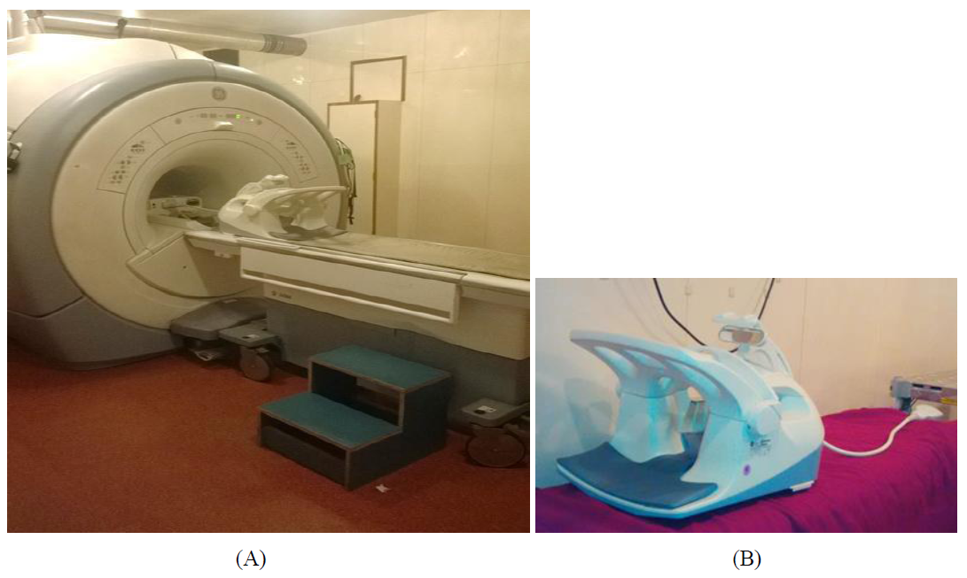
Data is collected in the year of 2018-2019 from the department of radiology, Chettinad Hospital and Research Institute (CHRI). Hospital-based descriptive and prospective study. Twenty-five patients were included in this study, selected randomly based on the patient’s history of trauma and non- traumatic symptoms of high blood pressure, sudden & severe headache, loss of balance or coordination and patients with hemorrhage in the brain who undergone MRI were included. Patients with metallic implants and cardiac pacemakers, claustrophobic patients were excluded. We used 1.5 Tesla SIGNA GE HDxt MRI scanner (Figure 1 A) with an 8 channel NV (NAVIGATOR) radiofrequency coil (Figure 1B). The bore size of the machine is 60cm. Both GRE and SWI sequence were used for recording the brain image. The number of patients were further classified into three subcategories based on their clinical history as follows, those with traumatic brain injury (TBI), with hypertention (H-BP) and with amyloids angiopathy (AA).

Figure 1: (A) 1.5 T MRI Scanner; (B) 8 Channel Head Coil.
3. Data Analysis
The research proposal was approved by the institutional human ethical committee of the Chettinad Academy of Research and Education (IHEC-CARE). The axial section of the brain image of SWI and GRE images which shows haemorrhage with hypointense signal for all the 25 patients were analyzed using ImageJ (NIH software package) and the area of hemorrhage was marked manually in the work station and also automatically selected by using Image package using thresholding and binarising technique. The computed
areas were used further for descriptive analysis using SPSS software. Table 1 shows the MR acquisition parameters for both the sequence.
Step: 1
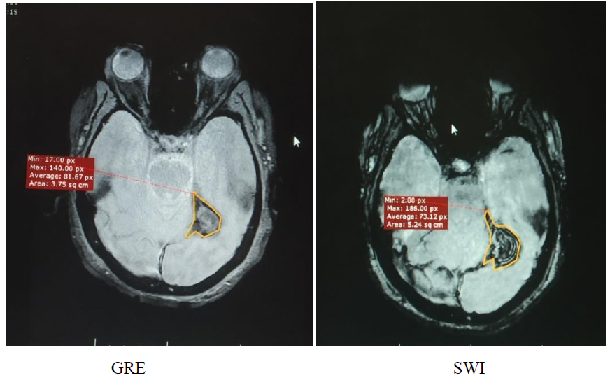
Areas are measured from the slices which show greater hemorrhages in GRE and SWI sequence. The areas of hemorrhage are manually drawn and calculated in sq.cm (Figure 2).
Step: 2
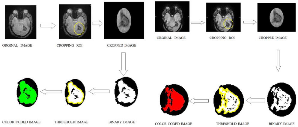
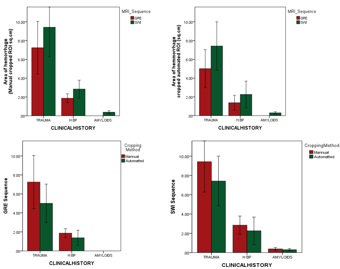
Areas are measured from the slices which show greater hemorrhages in GRE and SWI sequence. The areas of hemorrhage are automatically measured and calculated. Using ImageJ software the areas of hemorrhage are cropped and converted into the binary image and then thresholded to measure the area of bleed (Figure 3). The area of measurements is calibrated with MRI work station and converted to square centimetres (sq. Cm). The values of the measured area are expressed as mean and standard deviation in both manual and automated methods and compared using a paired t-test (Table 2). The calculated mean area and standard deviation are presented a bar chart with whiskers (Figure 4).
|
MR acquisition Parameter |
GRE |
SWI |
|
Plane |
Axial |
Axial |
|
Field of View (FOV) mm |
24 |
24 |
|
Repetition Time (TR) msec |
500 |
62 |
|
Echo Time (TE) msec |
15 |
41 |
|
Matrix |
512 x 224 |
352 x 224 |
|
Fatsat |
No |
No |
|
Slice thickness (mm) |
5 |
3 |
|
Inter slice (mm) |
1.5 |
- |
|
NEX |
1 |
1 |
|
Timing (min) |
1.28 |
3.28 |
|
IV contrast |
No |
No |
Table 1: MR acquisition parameters for GRE and SWI sequence.

Figure 2: Manual Measurement of Hemorrhage.
|
Clinical conditions |
Haemorrhage detected% GRE |
Haemorrhage detected% SWI |
Area of haemorrhage |
||||
|
Manual (ROI) sq.cm |
Automated (ROI) sq.cm |
||||||
|
GRE |
SWI |
GRE |
SWI |
||||
|
TBI |
16/17=92% |
17/17=100% |
7.70 ± 5.64 |
5.32 ± 4.09 |
9.42 ± 6.49 |
7.42 ±5.32 |
|
|
H-BP |
4/4=100% |
4/4=100% |
1.73 ± 0.47 |
1.21 ± 0.79 |
2.71 ± 0.94 |
1.84 ±1.42 |
|
|
AA |
0/4=0% |
4/4=100% |
- |
- |
0.385 ± 0.152 |
AA |
|
Table 2: Clinical conditions and calculated haemorrhage area are presented.

Figure 3: GRE and SWI -Process Of Areas Measurements.

Figure 4: Mean area of hemorrhage computed manually and by automated methods.
4. Discussion
In this study, patients with brain haemorrhage were included and the qualitative assessment was done for the presence of blooming by a radiologist. The superiority of SWI comparing to GRE is evidenced in our preliminary observation. However, SWI sequences take more time to acquire than simple T2*, and may sometimes the image quality is not due to motion artifacts. Further to this, possibly more false-positive and ambiguous lesions may also occur with SWI sequence and are more difficult to interpret as it comprises many dark structures that could be misguided for microbleeds [8]. Earlier studies suggest that cerebral microbleeds (CMBs) can be identified better on SWI than GRE [8]. An earlier study with 14 patients found that SWI acquired using 3T were associated with better- identified microbleeds and showed a good contrast-to-noise ratio (CNR) [9]. Another study on 141 patients with dementia, CMBs were detected more commonly on SWI compared with GRE [10]. In the present study, if lesions are present, the areas are measured in quantitative manner by both manual and automatic methods with ImageJ software. In which, the areas of haemorrhage were cropped and converted into thresholded and made to a binary image and measured automatically and calibrated to sq.cm. The values of manual and automated methods are further analysed and grouped to mean and standard deviation based on the clinical observation, such as traumatic brain injury, high blood pressure and amyloid haemorrhages. Apart from the advantages associated with SWI, there are limitations some concerned with SWI, such as there is no gold standard view for haemorrhages to validate its reliability in MRI [11]. There will be little confusion in the detection of calcifications [12]. Considering the above limitations, in this study, we observed that 1) SWI sequence is more sensitive in detecting CH when compared to GRE, further to this 2) automated ROI selection improved the quantifying the area of lesion in the MR image.
5. Conclusion
In this prospective study, 25 patients have been included with a brain hemorrhage and have performed a qualitative assessment for the presence of blooming. If present, areas are measured in a quantitative manner by both manual and automatic methods. In this study, we showed that SWI has a higher sensitivity for micro hemorrhage detection when compared to GRE. Additionally, we found that the sensitivity and reliability of hemorrhage detection using automated area measurement methods is high when compared to manual area measurement methods.
References
- Brouwers H B, Goldstein J N. Therapeutic strategies in acute intracerebral hemorrhage. Neurotherapeutics : the journal of the American Society for Experimental NeuroTherapeutics 9 (2012): 87-98.
- Kidwell C S, Chalela J A, Saver J L, et al. Comparison of MRI and CT for Detection of Acute Intracerebral Hemorrhage. JAMA 292 (2004): 1823-1830.
- Vymazal J, Rulseh A M, Keller J, et al. Comparison of CT and MR imaging in ischemic stroke. Insights Imaging 3 (2012): 619-627.
- Brazzelli M, Sandercock P A G, Chappell F M, et al. Magnetic resonance imaging versus computed tomography for detection of acute vascular lesions in patients presenting with stroke symptoms. Cochrane Database of Systematic Reviews (2009).
- Kummer R v. MRI: The New Gold Standard for Detecting Brain Hemorrhage? Stroke 33 (2002): 1748-1749.
- Lin M D D, Filippi C G, Steever A B, et al. Detection of Intracranial Hemorrhage: Comparison between Gradient-echo Images and b0 Images Obtained from Diffusion-weighted Echo-planar Sequences. American Journal of Neuroradiology 22 (2001): 1275-1281.
- Halefoglu A M, Yousem D M. Susceptibility weighted imaging: Clinical applications and future directions. World J Radiol 10 (2018): 30-45.
- Cheng A-L, Batool S, McCreary Cheryl R, et al. Susceptibility-Weighted Imaging is More Reliable Than T2*-Weighted Gradient-Recalled Echo MRI for Detecting Microbleeds. Stroke 44 (2013): 2782-2786.
- Nandigam R N, Viswanathan A, Delgado P, et al. MR imaging detection of cerebral microbleeds: effect of susceptibility-weighted imaging, section thickness, and field strength. AJNR. American journal of neuroradiology 30 (2009): 338-343.
- Goos J D, van der Flier W M, Knol D L, et al. Clinical relevance of improved microbleed detection by susceptibility-weighted magnetic resonance imaging. Stroke 42 (2011): 1894-1900.
- Romanova A L, Nemeth A J, Berman M D, et al. Magnetic resonance imaging versus computed tomography for identification and quantification of intraventricular hemorrhage. J Stroke Cerebrovasc Dis 23 (2014): 2036-2040.
- Hingwala D, Kesavadas C, Thomas B, et al. Clinical utility of susceptibility-weighted imaging in vascular diseases of the brain. Neurology India 58 (2010): 602-607.
