Giant Malignant Phyllodes Tumor of the Breast
Article Information
atarina Abreu Silva*, Madalena Souto Moura, Mariana Gomes Afonso, Susana Lopes Rodrigues, Ana Catarina Silva
Hospital Pedro Hispano, Portugal
*Corresponding Author: Dr. Catarina Abreu Silva, Hospital Pedro Hispano, Portugal
Received: 27 July 2020; Accepted: 25 August 2020; Published: 01 September 2020
Citation: Catarina Abreu Silva, Madalena Souto Moura, Mariana Gomes Afonso, Susana Lopes Rodrigues, Ana Catarina Silva. Giant Malignant Phyllodes Tumor of the Breast. Journal of Radiology and Clinical Imaging 3 (2020): 085-088.
Share at FacebookAbstract
Phyllodes tumor is a rare fibroepithelial tumor of the breast that account for less than 1% of all breast neoplasms [1]. Clinically, it is typically a large, smooth and fast-growing mass that forms from the periductal stroma of the breast [2]. Phyllodes tumor can be considered benign, borderline, or malignant and the percentage of malignant tumors ranges from 23 to 50%. This classification depends on histologic features, as the degree of stromal cellularity and atypia, mitotic count and stromal overgrowth [3]. We report a case of a huge malignant phyllodes tumor in a 62-year old woman who presented with a large breast mass with 10-month disease evolution time. The patient underwent total mastectomy and the pathology analysis revealed a malignant tumor.
Keywords
Breast neoplasms; Phyllode tumor; Breast ultrasonography; Radical mastectomy
Breast neoplasms articles; Phyllode tumor articles; Breast ultrasonography articles; Radical mastectomy articles
Breast neoplasms articles Breast neoplasms Research articles Breast neoplasms review articles Breast neoplasms PubMed articles Breast neoplasms PubMed Central articles Breast neoplasms 2023 articles Breast neoplasms 2024 articles Breast neoplasms Scopus articles Breast neoplasms impact factor journals Breast neoplasms Scopus journals Breast neoplasms PubMed journals Breast neoplasms medical journals Breast neoplasms free journals Breast neoplasms best journals Breast neoplasms top journals Breast neoplasms free medical journals Breast neoplasms famous journals Breast neoplasms Google Scholar indexed journals Phyllode tumor articles Phyllode tumor Research articles Phyllode tumor review articles Phyllode tumor PubMed articles Phyllode tumor PubMed Central articles Phyllode tumor 2023 articles Phyllode tumor 2024 articles Phyllode tumor Scopus articles Phyllode tumor impact factor journals Phyllode tumor Scopus journals Phyllode tumor PubMed journals Phyllode tumor medical journals Phyllode tumor free journals Phyllode tumor best journals Phyllode tumor top journals Phyllode tumor free medical journals Phyllode tumor famous journals Phyllode tumor Google Scholar indexed journals Breast ultrasonography articles Breast ultrasonography Research articles Breast ultrasonography review articles Breast ultrasonography PubMed articles Breast ultrasonography PubMed Central articles Breast ultrasonography 2023 articles Breast ultrasonography 2024 articles Breast ultrasonography Scopus articles Breast ultrasonography impact factor journals Breast ultrasonography Scopus journals Breast ultrasonography PubMed journals Breast ultrasonography medical journals Breast ultrasonography free journals Breast ultrasonography best journals Breast ultrasonography top journals Breast ultrasonography free medical journals Breast ultrasonography famous journals Breast ultrasonography Google Scholar indexed journals Radical mastectomy articles Radical mastectomy Research articles Radical mastectomy review articles Radical mastectomy PubMed articles Radical mastectomy PubMed Central articles Radical mastectomy 2023 articles Radical mastectomy 2024 articles Radical mastectomy Scopus articles Radical mastectomy impact factor journals Radical mastectomy Scopus journals Radical mastectomy PubMed journals Radical mastectomy medical journals Radical mastectomy free journals Radical mastectomy best journals Radical mastectomy top journals Radical mastectomy free medical journals Radical mastectomy famous journals Radical mastectomy Google Scholar indexed journals mastectomy articles mastectomy Research articles mastectomy review articles mastectomy PubMed articles mastectomy PubMed Central articles mastectomy 2023 articles mastectomy 2024 articles mastectomy Scopus articles mastectomy impact factor journals mastectomy Scopus journals mastectomy PubMed journals mastectomy medical journals mastectomy free journals mastectomy best journals mastectomy top journals mastectomy free medical journals mastectomy famous journals mastectomy Google Scholar indexed journals axillary lymphadenopathy articles axillary lymphadenopathy Research articles axillary lymphadenopathy review articles axillary lymphadenopathy PubMed articles axillary lymphadenopathy PubMed Central articles axillary lymphadenopathy 2023 articles axillary lymphadenopathy 2024 articles axillary lymphadenopathy Scopus articles axillary lymphadenopathy impact factor journals axillary lymphadenopathy Scopus journals axillary lymphadenopathy PubMed journals axillary lymphadenopathy medical journals axillary lymphadenopathy free journals axillary lymphadenopathy best journals axillary lymphadenopathy top journals axillary lymphadenopathy free medical journals axillary lymphadenopathy famous journals axillary lymphadenopathy Google Scholar indexed journals cutaneous involvement articles cutaneous involvement Research articles cutaneous involvement review articles cutaneous involvement PubMed articles cutaneous involvement PubMed Central articles cutaneous involvement 2023 articles cutaneous involvement 2024 articles cutaneous involvement Scopus articles cutaneous involvement impact factor journals cutaneous involvement Scopus journals cutaneous involvement PubMed journals cutaneous involvement medical journals cutaneous involvement free journals cutaneous involvement best journals cutaneous involvement top journals cutaneous involvement free medical journals cutaneous involvement famous journals cutaneous involvement Google Scholar indexed journals Doppler evaluation articles Doppler evaluation Research articles Doppler evaluation review articles Doppler evaluation PubMed articles Doppler evaluation PubMed Central articles Doppler evaluation 2023 articles Doppler evaluation 2024 articles Doppler evaluation Scopus articles Doppler evaluation impact factor journals Doppler evaluation Scopus journals Doppler evaluation PubMed journals Doppler evaluation medical journals Doppler evaluation free journals Doppler evaluation best journals Doppler evaluation top journals Doppler evaluation free medical journals Doppler evaluation famous journals Doppler evaluation Google Scholar indexed journals hypercellular stroma articles hypercellular stroma Research articles hypercellular stroma review articles hypercellular stroma PubMed articles hypercellular stroma PubMed Central articles hypercellular stroma 2023 articles hypercellular stroma 2024 articles hypercellular stroma Scopus articles hypercellular stroma impact factor journals hypercellular stroma Scopus journals hypercellular stroma PubMed journals hypercellular stroma medical journals hypercellular stroma free journals hypercellular stroma best journals hypercellular stroma top journals hypercellular stroma free medical journals hypercellular stroma famous journals hypercellular stroma Google Scholar indexed journals local radiotherapy articles local radiotherapy Research articles local radiotherapy review articles local radiotherapy PubMed articles local radiotherapy PubMed Central articles local radiotherapy 2023 articles local radiotherapy 2024 articles local radiotherapy Scopus articles local radiotherapy impact factor journals local radiotherapy Scopus journals local radiotherapy PubMed journals local radiotherapy medical journals local radiotherapy free journals local radiotherapy best journals local radiotherapy top journals local radiotherapy free medical journals local radiotherapy famous journals local radiotherapy Google Scholar indexed journals
Article Details
1. Case Report
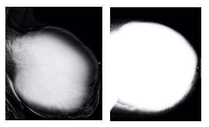
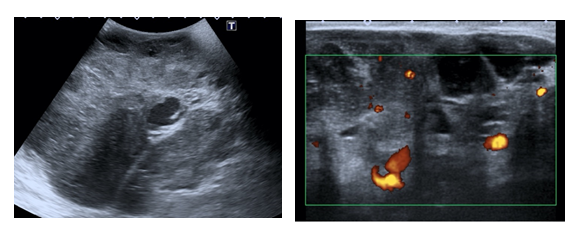
A 62-year old woman presented with a large breast mass with 10-month disease evolution time on left breast, with accelerated growth in recent months. The patient had no known breast disease or family history of breast cancer. Physical exam reveals a protruding giant mass occupying all left breast but with no evident signs of cutaneous involvement or axillary lymphadenopathy. The patient was submitted to mammography and breast ultrasound to a better characterization of the breast mass. Mammogram revealed a huge dense mass obscuring all normal breast tissue (Figure 1) and ultrasonography showed a volumous solid mass with heterogeneous echotexture, with endoluminal cystic areas and internal flow with Doppler evaluation (Figure 2). The patient was submitted to ultrasound-guided biopsy that reveals a phyllodes tumor and then underwent total mastectomy.
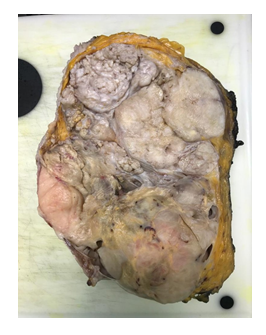
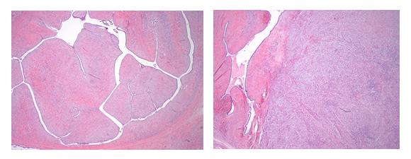
A breast mass with expansive limits and heterogeneous appearance, measuring 26.5 × 26 cm in dimensions, (Figure 3) was resected and the microscopic analysis showed a lesion with fibro-adenomatous changes with proliferation of epithelial and stromal components. The hypercellular stroma demonstrated marked nuclear pleomorphism, increased mitotic activity as well as irregular large cystic spaces with protruding leaf-like structures and areas of necrosis and hemorrhage (Figure 4). These features were consistent with malignant phyllodes tumor of the breast. Postoperatively the patient received local radiotherapy and is now on follow-up without signs of distant metastasis.

Figure 1: Mammography demonstrated a huge well circumscribed hyperdense mass encompassing almost the entire left breast. No signs of suspicion microcalcifications are seen.

Figure 2: Ultrasound of the left breast showed a heterogeneous mass, difficult to accurately measure given it extent. Some cystic spaces are seen as well as some solid areas with vascularity.

Figure 3: Left breast almost totally occupied by a heterogeneous multilobulated neoplasia, with foci of leaf-like appearance, extensive fleshy solid areas, necrosis and focally infiltrative borders.

Figure 4: A. Biphasic fibroepithelial neoplasia with leaf-like stromal fronds capped by epithelial cells without atypia (2x). B. Extensive areas presenting all the criteria required for a diagnosis of malignant phyllodes tumor: stromal overgrowth, increased stromal cellularity, marked stromal nuclear pleomorfism, mitotic activity, necrosis (not showed) and infiltrative borders (not showed) (2x).
2. Discussion
Phyllodes tumor is a rare breast tumor that occurs more frequently in women between 35-55 years old and has an incidence of less than 1% of all breast neoplasms. Clinically, these tumors frequently present as a rapid growth mass and they’re usually painless and difficult to differentiate from fibroadenomas [1, 2]. Histologically, phyllodes tumors are biphasic neoplasms characterized by the presence of stromal and epithelial components with a leaf-like appearance and cystic spaces oh histological examination. The stromal elements project into the ductal lumen with a foliaceous aspect. Phyllodes tumors are classified into benign, borderline or malignant tumors depending on histological characteristics as increased mitotic activity, degree of cellular atypia, and stromal proliferation [2-4].
Radiologically, there are no mammographic or ultrasonographic pathognomonic findings but these tumors usually present as isodense and well-circumscribed masses on mammogram with solid and cystic areas and internal flow on ultrasound with Doppler evaluation [2, 4]. Complete surgical resection of these tumors with a wide local excision is the treatment of choice as there is a high tendency to recurrences, in up to 25% of the cases. For borderline and malignant phyllodes tumors or in cases of positive margins or tumor recurrence, mastectomy is the first option and it’s essential to follow up the patients due to the risk of distant metastasis (up to 10%) [5-7].
3. Conclusion
In conclusion, it’s important that clinicians, radiologists and pathologists be familiar with the features of phyllodes tumors as they are rare breast tumors with possibility of malignancy and rapid growth [1, 2].
References
- Tan H, Zhang S, Liu H, et al. Imaging findings in phyllodes tumors of the breast. 2011.Feder JM, De paredes ES, Hogge JP, et al. Unusual breast lesions: radiologic-pathologic correlation. Radiographics 19 (1999): S11-S26.
- Yabuuchi H, Soeda H, Matsuo Y, et al. Phyllodes tumor of the breast: correlation between MR findings and histologic grade. Radiology 241 (2006): 702-709.
- Buchberger W, Strasser K, Heim K, et al. Phylloides tumor: findings on mammography, sonography, and aspiration cytology in 10 cases. AJR Am J Roentgenol 157 (1991): 715-719.
- Liang MI, Ramaswamy B, Patterson CC, et al. Giant breast tumors: surgical management of phyllodes tumors, potential for reconstructive surgery and a review of literature. World J Surg Oncol 6 (2008): 117.
- Yohe S, Yeh IT. “Missed” diagnosis of Phyllodes tumor on breast biopsy: pathological clues to its recognition. Int J Surg Pathol 16 (2008): 137-142.
- Reinfuss M, Mitus J, Duda K, et al. The treatment and prognosis of patients with phyloides tumour of the breast: an analysis of 170 cases. Cancer 77 (1996): 910-916.
