Evaluation of Dependence between Left Atrial Appendage Flow Velocity Examined in Transoesophageal Echocardiography (TTE) and CHA2DS2-Vasc Score in Patients with Atrial Fibrillation (AF)
Article Information
Jaroslaw Szponar1,2, Anna Krajewska1,2, Michal Tchórz1,2, Piotr Danielewicz2, Anna Kujawa2, Agnieszka Górska1,2, Tomasz Slomka3
1Toxicology Clinic, Medical University of Lublinal. Krasnicka 100, 20-718, Lublin, Poland
2Clinical Department of Toxicology and Cardiology, Cardinal Stefan Wyszynski Regional Specialist Hospital, Al. Krasnicka 100, 20-718 Lublin, Poland
3Department of Medical Informatics and Statistics with E-learning Lab, Medical University of Lublin, Poland
*Corresponding author: Jaroslaw Szponar, Stefan Wyszynski Regional Specialist Hospital, Al. Krasnicka 100, 20-718 Lublin, Poland.
Received: 25 January 2022; Accepted: 03 February 2022; Published: 15 February 2022
Citation: Jaroslaw Szponar, Anna Krajewska, Michal Tchórz, Piotr Danielewicz, Anna Kujawa, Agnieszka Górska, Tomasz Slomka. Evaluation of Dependence between Left Atrial Appendage Flow Velocity Examined in Transoesophageal Echocardiography (TTE) And CHA2DS2-Vasc Score in Patients With Atrial Fibrillation (AF). Archives of Clinical and Biomedical Research 6 (2022): 240-247.
Share at FacebookAbstract
Objectives: The objective of the paper was evaluation of link between 1) left atrial appendage flow velocity (LAA) in patients with Atrial Fibrillation (AF) examined during transoesophageal echocardiography and 2) CHA2DS2-VASc score obtained by the patient.
Methods and results: There were 450 people examined (N=450). Transoesophageal echocardiography with doppler sample placed 1 centimetre below LAA ostium was performed in all the patients. Pulse Wave doppler technique (PW) was used. It was determined that there is a key dependence between examined features (p=0,012). The content is weak and negative though. Left atrial appendage flow Velocity (V) is expected to be lower for each next class.
Conclusions: The dependence between two parameters (the left atrial appendage flow velocity and the points in CHA2DS2-VASc score) was indicated. In future it may be possible to use the left atrial appendage flow velocity as a new factor qualifying patients for oral anticoagulation therapy.
Keywords
Atrial fibrillation (AF); CHA2DS2-Vasc Score; Left atrial appendage (LAA)
Atrial fibrillation (AF) articles; CHA2DS2-Vasc Score articles; Left atrial appendage (LAA) articles
Article Details
1. Introduction
In Poland around 600.000-700.000 people suffer from atrial fibrillation. The occurrence increases along with age. In the age group <50 it affects 0,15 of population, >75 it occurs in around 10% of population and increases to around 18% of affected after the age of 85 [1]. The frequency of AF occurrence changes dependently on age and sex. AF is present in 0,12- 0,16 % in the age below 49, 3,7-4,2 % in the age of 60-70 and 10-17% for people in the age of 80 and older [2-5]. AF occurs more often in men than in women (frequency indicator is 1,2:1). Despite higher frequency in men, women are majority of patients suffering from AF due to their longevity [6-10]. The main clinical issue, besides symptoms linked to arrythmia are its complications. Framingham examination indicated that the risk of stroke is 17 times higher in patients with rheumatic valvular disease, the risk of thromboembolism is 5 times higher in patients with nonvalvular atrial fibrillation, there is 2-3 times increase in occurrence of symptomatic congestive heart failure and the frequency of hospitalisation as well as 2 times increase in deaths. The most frequent locations of thromboembolism, besides ischaemic stroke, are: renal artery, mesenteric artery, upper and lower limbs arteries and arteries around pelvic organs. The most frequent complication of atrial fibrillation is stroke which is characterised by worse course and prognosis in comparison to stroke with different etiology. It was proven that the risk of next stroke in patients with AF is 4 times higher and the risk of death is 2 times higher. Atrial fibrillation is confirmed in 15,6% patients with stroke whereas the diagnosis in the half on them is based on a routine electrocardiography [11]. According to data provided, among the most essential therapies in AF patients are not only rhythm-control and rate-control strategy but also preventing thromboembolism by using oral anticoagulation therapy.
1.1 Etiopathogenesis of thromboembolism incidents
In etiopathogenesis of increased thromboembolism risk in AF patients all 3 factors described in Virchow's triad are involved. In patients with AF there is both, significant lower blood flow and vascular wall disfunction (atrium in this case) as well as increased blood coagulability. In consequence such factors result in increased risk of embolism. Very fast chaotic atrium systole, typical to AF, leads to significant decrease of blood flow within atriums and in Left Atrial Appendage (LAA). This causes the spontaneus echo contrast phenomenon observed in echocardiographic examination. In patients with AF increased plasma concentration of prothrombotic factors such as: fibrinogen and d-dimer was observed. It proves that those patients had higher blood coagulability. Overexpression of von Willebrand factor and tissue factor in atriums endocardia is also observed in patients with AF with thromboembolism. It is mainly activation of coagulation system that is responsible for thromboembolism during AF in atria, in contrary to arterial bed where dominant role is played by platelets. This different mechanism of creating embolic material has significant clinical importance. It explains higher effectiveness of oral anticoagulants rather than antiplatelet therapy to prevent thromboembolism in patients with AF [12,13].
1.2 Risk factors of thromboembolism
The risk of stroke in patients with AF, just as in all populations, increases along with age. Moreover, it was stated that in patients with AF left atrium is enlarged and left atrial appendage flow velocity is slower along with age. Such phenomenon strongly predisposes to formation of thromboembolic material. Arterial hypertension intercurrent with AF is also linked to lower blood flow in left atrium and it fosters the spontaneus echo contrast phenomenon in the left atrial appendage. Arterial hypertension increases the risk of stroke in patients with AF two times. Diabetes can predispose to formation of thromboembolic material by increased concentration of substances with prothrombotic features in blood. Based on available data it is estimated that diabetes increases the risk of stroke 1,5-2 times in patients with AF and diabetes. Chronic heart failure can also significantly increase proclivity to formation of thromboembolic material. Patients with heart failure have significantly increased concentration of von-Willebrand factor, dissoluble thrombomodulin and E-selectine. In SPAF trial heart failure was independent risk factor of thromboembolism complications in patients with AF (7%/year) [13,14]. Stroke is the most frequent complication of AF. Stroke/TIA is also the strongest independent risk factor of next thromboembolic incident occurrence, increasing the risk of next stroke 5 times. On one hand, stroke is a consequence of prothrombotic condition, whereas on the other hand it is caused by increased predisposition to forming thromboembolic material in patients with AF resulting from balance between pro-anticoagulation factors [14].
1.3 Risk factors of thromboembolism evaluation
The risk of thromboembolic incident occurrence in patients with AF is not homogenous and is dependant on age, sex and other intercurrent diseases mentioned in previous chapter of the paper. Scientists from ,,The Stroke In Atrial Fibrillation” task force included prior stroke/ TIA (RR –relative risk 2,5), old age (RR 1,5/10years), arterial hypertension (RR 2,0), diabetes (RR1,8), female sex (RR 1,6) to stroke risk factors. Based on very similar clinical parameters members of Scientific Project SPAF (Stroke Prevention In Atrial Fibrillation) created widely known point scale for stroke risk evaluation - CHADS2 Score (Congestive Heart Failure, Hypertension, Age>75, Diabetes mellitus, Stroke/TIA). Each parameter has 1 point assigned in this Score. Only prior stoke/transient ischemic attack has 2 points assigned. The Score, however, does not enable isolating very low stroke risk group. What is more, its positive predictive value in stroke evaluation is 0,58. Extended Score CHA2DS2-VASc (Table 1) increases its positive predictive value and allows strong predictive negative value [14-16]. CHA2DS2-VASc Score has many advantages. It is simple and based mainly on information gathered during the interview and physical examination of the patient. Is was also validated.
|
CHA2DS2-VASc Score |
Condition |
Points |
|
C-congestive heart failure/LV dysfunction |
Congestive heart failure (or Left ventricular systolic dysfunction) |
1 |
|
H - hypertension |
Hypertension |
1 |
|
A-age |
Age≥75 |
2 |
|
Diabetes mellitus |
Diabetes |
1 |
|
Stroke/TIA/Thromboembolism History |
Prior stroke/TIA /Thromboembolism |
2 |
|
V - vascular disease history |
Vascular disease, peripheral artery disease, myocardial infraction, aortic plaque |
1 |
|
A-age |
Age 65-74 |
1 |
|
Sc-sex category |
Female sex |
1 |
Table 1: CHA2DS2-VASc Score
Estimated risk of stroke in CHADS VASc Score was presented in (Table 2) [16].
|
Points in CHA2DS2-VASc Score |
Number of examined patients |
Percentage of stroke by age (%/year) |
|
0 |
1 |
0% |
|
1 |
422 |
1,3% |
|
2 |
1230 |
2,2% |
|
3 |
1730 |
3,2% |
|
4 |
1718 |
4,0% |
|
5 |
1159 |
6,7% |
|
6 |
679 |
9,8% |
|
7 |
294 |
9,6% |
|
8 |
82 |
6.7% |
|
9 |
14 |
15,2% |
Table 2: Estimated risk of stroke based on CHA2DS2-VASc Score.
Stroke risk evaluation with this Score enables identification of low risk patients as well as patients that can benefit more from oral anticoagulation. On the other hand, oral anticoagulation therapy is linked to increased risk of bleedings, hemorrhagic stroke included. It is important to be advised that guidelines for oral anticoagulation therapy and the choice of medication are not unambiguous for patients burdened with medium stroke risk or with high probability of haemorrhage. In such patients, evaluation of risk factors in echocardiography can be useful because the presence of such factors will indicate necessity of inclusion chronic oral anticoagulant therapy (Table 3) [17,18].
|
Echocardiographic Risk Factors of Embolism in Artial Fibrillation |
|
EF< 35% |
|
Thick, spontaneous contrast in left atrial appendage |
|
Left atrial appendage thrombus |
|
Left atrial appendage disfunction (blood flow below 20cm/s) |
|
Complex aortic plaque |
Table 3: Echocardiographic factors of embolism in atrial fibrillation
According to Virchow, by interfering blood flow, atrial fibrillation fosters creation of thrombus inside atriums – most frequently in left atrial appendage (LAA). The most significant is the left atrial appendage shape, that is usually very complex, as well as its satellite location against left atrium. These are additional factors for thrombus creation especially in situations with impairment of mechanical activity, like in arrhythmia. Due to high sensitivity and specificity transoesophageal echocardiography is considered a golden standard in detecting embolic material in LAA as well as symptoms of atrial appendage disfunction fostering thrombus creation [18-21]. Those include: spontaneous contrast in left atrial appendage and left atrial appendage flow velocity (evaluated by pulse wave doppler technics) below 20cm/s [22-25]
2. Objectives of This Work
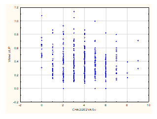
The subject of this trial was to determine dependence between the number of points obtained in CHA2DS2-VASc Score and the left atrial appendage flow velocity marked in transoesophageal echocardiography.
3. Materials and Methods
There were 450 persons examined (195 women, 255men) with atrial fibrillation. Average age of patients was 71. Each patient was evaluated in CHA2DS2-VASc Score receiving 0-9 points. Average points were 3,6. Transoesophageal echocardiography with doppler sample placed 1 centimetre below LAA ostium was performed in all the patients. Pulse wave doppler technics (PW) was used. The measurement was done by setting doppler gate collaterally to blood flow in 1/3 closer part of atrial appendage around 1 centimetre below ostium [22-26]. When LLA function was maintained, the rates were recorded as oscillatory above and below isoelectric line. When there was systolic atrial appendage disfunction, lower values were recorded. According to available bibliography, recorded rates velocities below 20 cm/s indicate atrial appendage disfunction fostering thrombus creation [26-28].
In this paper, the dependence between the number of points obtained in CHA2DS2-VASc Score and lowered left atrial appendage flow velocity was evaluated.
4. Results
Nonparametric test of Spearman rank
Statistical significance Alpha = 0,05
|
|
Spearmna;s Rank correlation (Sheet2) BD deleted by pairs Correlation coefficients are significant when p<.050000 |
||||||
|
|
|
|
||||
|
450 |
-0.118730 |
-2.53095 |
0.011717 |
|||
It was stated that there is statistically significant dependence between examined features (p=0,012). This dependence is weak and negative though. For each next class, the expected V value of left atrial appendage flow velocity is lower.
4. Conclusions
Obtained results proved correlation between the left atrial appendage flow velocity and points obtained in CHA2DS2-VASc Score. Therefore, the evaluation of left atrial appendage flow velocity in transoesophageal echocardiography examination could potentially be used as individual or supportive parameter in qualifying patients for oral anticoagulation therapy in the future.
References
- Mizia Stec K. Trusz- Gluza M. Arrythmias. Medical Tribune 2018.
- Kirchhof P, Benussi S, Kotecha D, et al. ESC Guidelines for the management of atrial fibrillation developed in collaboration with EACTS European Heart Journal 37 (2016): 2893-2962,
- Go A , Hylek E, Phillips K, et al. Prevalence of Diagnosed Atrial Fibrillation in Adults: National Implications for Rhythm Management and Stroke Prevention: the AnTicoagulation and Risk Factors In Atrial Fibrillation (ATRIA) Study. JAMA 285 (2001): 2370-2375.
- Zoni-Berisso M, Lercari F, Carazza T, et al. Epidemiology of atrial fibrillation: European perspective. Clin Epidemiol 6 (2014): 213-220.
- Fitzmaurice DA, Hobbs FD, Jowett S, et al. Screening versus routine practice in detection of atrial fibrillation in patients aged 65 or over: cluster randomised controlled trial. BMJ 335 (2007): 383.
- Rho RW, Page RL. Asymptomatic atrial fibrillation. Prog Cardiovasc Dis 48 (2005): 79-87.
- Chugh SS, Havmoeller R, Narayanan K, et al. Worldwide epidemiology of atrial fibrillation: a Global Burden of Disease 2010 Study. Circulation 129 (2014): 837-847
- Wilke T, Groth A, Mueller S, et al. Incidence and prevalence of atrial fibrillation: an analysis based on 8.3 million patients. Europace 15 (2013): 486-493.
- Fang MC, Singer DE, Chang Y, et al. Gender differences in the risk of ischemic stroke and peripheral embolism in atrial fibrillation: the AnTicoagulation and Risk factors In Atrial fibrillation (ATRIA) study. Circulation 112 (2005): 1687-1691.
- Piccini JP, Hammill BG, Sinner MF, et al. Incidence and prevalence of atrial fibrillation and associated mortality among Medicare beneficiaries, 1993-2007.Circ Cardiovasc Qual Outcomes 5 (2012):85-93.
- Schnabel RB, Yin X, Gona P, et al. 50 year trends in atrial fibrillation prevalence, incidence, risk factors, and mortality in the Framingham Heart Study: a cohort study. Lancet J 386 (2015):154-162.
- Watson T, Shantsila E, Lip GY. Mechanisms of thrombogenesis in atrial fibrillation: Virchow's triad revisited. Lancet 373 (2009): 155-166.
- Lip GY, Nieuwlaat R, Pisters R, et al. Refining clinical risk stratification for predicting stroke and thromboembolism in atrial fibrillation using a novel risk factor-based approach: the euro heart survey on atrial fibrillation. Chest 137 (2010): 263-272.
- Goldman ME, Pearce LA, Hart RG, et al. J Am Soc Echocardiogr 12 (1999):1080-1087. Pathophysiologic correlates of thromboembolism in nonvalvular atrial fibrillation: I. Reduced flow velocity in the left atrial appendage (The Stroke Prevention in Atrial Fibrillation [SPAF-III] study).Circulation 112 (2005): 1687-1691.
- Wozakowska-Kaplon B. Atrial Fibrillation. Via Medica Gdansk (2016).
- Dluzniewski M, Syska-Suminska J, Kalarus Z. Atrial fibrillation-everyday therapy in general medicine. Wydawnictwo Czelej Lublin (2016).
- Kun Z, Lanlan S, Xinchun Y, et al. Correlation between cardiac rhythm, left atrial appendage flow velocity, and CHA2DS2-VASc score: Study based on transesophageal echo-cardiography and 2-dimensional speckle tracking. Clin Cardiol 40 (2017): 120-125.
- Transesophageal echocardiographic correlates of thromboembolism in high-risk patients with nonvalvular atrial fibrillation. The Stroke Prevention in Atrial Fibrillation Investigators Committee on Echocardiography. Ann Intern Med 128 (1998): 639-647.
- Clark CB, Telles Garcia NA, Hackett Renner C, et al. Correlation of Left Atrial Appendage Ejection Velocities with the CHADS2 and CHA2DS2-VASc Scores. Echocardiography (Mount Kisco, N.Y.) 33 (2016): 1195-2010
- Lee JM, Shim J, Uhm JS, et al. Impact of increased orifice size and decreased flow velocity of left atrial appendage on stroke in nonvalvular atrial fibrillation. Am J Cardiol 113 (2014): 963-969.
- Mugge A, Kuhn H, Nikutta P, et al. Assessment of left atrial appendage function by biplane transesophageal echocardiography in patients with nonrheumatic atrial fibrillation: identification of a subgroup of patients at increased embolic risk. J Am Coll Cardiol 23 (1994): 599-607.
- Fatkin D, Kelly RP, Feneley MP. Relations between left atrial appendage blood flow velocity, spontaneous echocardiographic contrast and thromboembolic risk in vivo. J Am Coll Cardiol 23 (1994): 961-969.
- Kortz RA, Delemarre BJ, van Dantzig JM, et al. Left atrial appendage blood flow determined by transesophageal echocardiography in healthy subjects. Am J Cardiol 71 (1993): 976-981
- Jue J, Winslow T, Fazio G, et al. Pulsed Doppler characterization of left atrial appendage flow. J Am Soc Echocardiogr 6 (1993): 237-244.
- Manabe K, Oki T, Tabata T. Transesophageal echocardiographic prediction of initially successful electrical cardioversion of isolated atrial fibrillation. Effects of left atrial appendage function. Jpn Heart J 38 (1997): 487-495.
- Donal E, Yamada H, Leclercq C, et al. The left atrial appendage, a small, blind-ended structure: a review of its echocardiographic evaluation and its clinical role. Chest 128 (2005): 1853-1862.
- Shah SJ, Bardo DM, Sugeng L, et al. Real-time three-dimensional transesophageal echocardiography of the left atrial appendage: initial experience in the clinical setting. J Am Soc Echocardiogr 21 (2008): 1362-1368
- Khurram IM, Dewire J, Mager M, et al. Relationship between left atrial appendage morphology and stroke in patients with atrial fibrillation. Heart Rhythm 10 (2013): 1843-1849.

