Early Diagnosis in Cutaneous Peripheral T-Cell Lymphoma Not Otherwise Specified
Article Information
Konstantinos Efthimiadis1, Julia Grammatikopoulou1, Despoina Noukari1, Aggeliki Georgiadou1, Ioannis Venizelos2, Christina Nikolaidou3, Evgenia Verrou4, Florentina Delli1*
1State Hospital for Skin and Venereal Diseases of Thessaloniki, Greece
2Consultant Histopathologist, Head of the Department, Hippokration General Hospital, Thessaloniki, Greece
3Consultant Histopthologist, Hippokration General Hospital, Thessaloniki, Greece
4Hematologist, Hematology Department Theagenio Cancer Center, Thessaloniki, Greece
*Corresponding Author: Florentina Delli, State Hospital for Skin and Venereal Diseases of Thessaloniki, Greece.
Received: 18 January 2022; Accepted: 27 January 2022; Published: 18 March 2022
Citation: Konstantinos Efthimiadis, Julia Grammatikopoulou, Despoina Noukari, Aggeliki Georgiadou, Ioannis Venizelos, Christina Nikolaidou, Evgenia Verrou, Florentina Delli. Early Diagnosis in Cutaneous Peripheral T-Cell Lymphoma Not Otherwise Specified. Archives of Clinical and Medical Case Reports 6 (2022): 203-208.
Share at FacebookKeywords
Cutaneous peripheral T-Cell lymphoma; T-cell antigens; Stem cell transplantation
Cutaneous peripheral T-Cell lymphoma articles; T-cell antigens articles; Stem cell transplantation articles
Covid-19 articles Covid-19 Research articles Covid-19 review articles Covid-19 PubMed articles Covid-19 PubMed Central articles Covid-19 2023 articles Covid-19 2024 articles Covid-19 Scopus articles Covid-19 impact factor journals Covid-19 Scopus journals Covid-19 PubMed journals Covid-19 medical journals Covid-19 free journals Covid-19 best journals Covid-19 top journals Covid-19 free medical journals Covid-19 famous journals Covid-19 Google Scholar indexed journals Cutaneous peripheral T-Cell lymphoma articles Cutaneous peripheral T-Cell lymphoma Research articles Cutaneous peripheral T-Cell lymphoma review articles Cutaneous peripheral T-Cell lymphoma PubMed articles Cutaneous peripheral T-Cell lymphoma PubMed Central articles Cutaneous peripheral T-Cell lymphoma 2023 articles Cutaneous peripheral T-Cell lymphoma 2024 articles Cutaneous peripheral T-Cell lymphoma Scopus articles Cutaneous peripheral T-Cell lymphoma impact factor journals Cutaneous peripheral T-Cell lymphoma Scopus journals Cutaneous peripheral T-Cell lymphoma PubMed journals Cutaneous peripheral T-Cell lymphoma medical journals Cutaneous peripheral T-Cell lymphoma free journals Cutaneous peripheral T-Cell lymphoma best journals Cutaneous peripheral T-Cell lymphoma top journals Cutaneous peripheral T-Cell lymphoma free medical journals Cutaneous peripheral T-Cell lymphoma famous journals Cutaneous peripheral T-Cell lymphoma Google Scholar indexed journals T-Cell lymphoma articles T-Cell lymphoma Research articles T-Cell lymphoma review articles T-Cell lymphoma PubMed articles T-Cell lymphoma PubMed Central articles T-Cell lymphoma 2023 articles T-Cell lymphoma 2024 articles T-Cell lymphoma Scopus articles T-Cell lymphoma impact factor journals T-Cell lymphoma Scopus journals T-Cell lymphoma PubMed journals T-Cell lymphoma medical journals T-Cell lymphoma free journals T-Cell lymphoma best journals T-Cell lymphoma top journals T-Cell lymphoma free medical journals T-Cell lymphoma famous journals T-Cell lymphoma Google Scholar indexed journals Computed tomography articles Computed tomography Research articles Computed tomography review articles Computed tomography PubMed articles Computed tomography PubMed Central articles Computed tomography 2023 articles Computed tomography 2024 articles Computed tomography Scopus articles Computed tomography impact factor journals Computed tomography Scopus journals Computed tomography PubMed journals Computed tomography medical journals Computed tomography free journals Computed tomography best journals Computed tomography top journals Computed tomography free medical journals Computed tomography famous journals Computed tomography Google Scholar indexed journals Ultra Sound articles Ultra Sound Research articles Ultra Sound review articles Ultra Sound PubMed articles Ultra Sound PubMed Central articles Ultra Sound 2023 articles Ultra Sound 2024 articles Ultra Sound Scopus articles Ultra Sound impact factor journals Ultra Sound Scopus journals Ultra Sound PubMed journals Ultra Sound medical journals Ultra Sound free journals Ultra Sound best journals Ultra Sound top journals Ultra Sound free medical journals Ultra Sound famous journals Ultra Sound Google Scholar indexed journals treatment articles treatment Research articles treatment review articles treatment PubMed articles treatment PubMed Central articles treatment 2023 articles treatment 2024 articles treatment Scopus articles treatment impact factor journals treatment Scopus journals treatment PubMed journals treatment medical journals treatment free journals treatment best journals treatment top journals treatment free medical journals treatment famous journals treatment Google Scholar indexed journals CT articles CT Research articles CT review articles CT PubMed articles CT PubMed Central articles CT 2023 articles CT 2024 articles CT Scopus articles CT impact factor journals CT Scopus journals CT PubMed journals CT medical journals CT free journals CT best journals CT top journals CT free medical journals CT famous journals CT Google Scholar indexed journals Radiology articles Radiology Research articles Radiology review articles Radiology PubMed articles Radiology PubMed Central articles Radiology 2023 articles Radiology 2024 articles Radiology Scopus articles Radiology impact factor journals Radiology Scopus journals Radiology PubMed journals Radiology medical journals Radiology free journals Radiology best journals Radiology top journals Radiology free medical journals Radiology famous journals Radiology Google Scholar indexed journals T-cell antigens articles T-cell antigens Research articles T-cell antigens review articles T-cell antigens PubMed articles T-cell antigens PubMed Central articles T-cell antigens 2023 articles T-cell antigens 2024 articles T-cell antigens Scopus articles T-cell antigens impact factor journals T-cell antigens Scopus journals T-cell antigens PubMed journals T-cell antigens medical journals T-cell antigens free journals T-cell antigens best journals T-cell antigens top journals T-cell antigens free medical journals T-cell antigens famous journals T-cell antigens Google Scholar indexed journals Stem cell transplantation articles Stem cell transplantation Research articles Stem cell transplantation review articles Stem cell transplantation PubMed articles Stem cell transplantation PubMed Central articles Stem cell transplantation 2023 articles Stem cell transplantation 2024 articles Stem cell transplantation Scopus articles Stem cell transplantation impact factor journals Stem cell transplantation Scopus journals Stem cell transplantation PubMed journals Stem cell transplantation medical journals Stem cell transplantation free journals Stem cell transplantation best journals Stem cell transplantation top journals Stem cell transplantation free medical journals Stem cell transplantation famous journals Stem cell transplantation Google Scholar indexed journals
Article Details
1. Case Report
Peripheral T-Cell Lymphomas (PTCLs) are uncommon and aggressive types of non-Hodgkin lymphoma. According to the 2016 WHO classification of Lymphoid Neoplasms, 29 discrete types of PTCL are recognized [1], typically divided into 4 categories, including: 1) disseminated or leukemic disease; 2) nodal disease; 3) extra-nodal disease; and 4) cutaneous disease. Among them, the most common entity is PTCL-NOS, accounting for 30% of all PTCLs [2]. Cutaneous PTCL-NOS (cPTCL-NOS) are defined as involving only the skin with no evidence of extra cutaneous disease at the initial staging or as cutaneous lesions preceding systemic involvement by 6 months. Fever, night sweats and unexplained weight loss are common in people with PTCL-NOS. It can occur at any age, but it is frequently observed in people in their 60s and affects men more than women. Considering that the majority of patients are at an advanced stage at the time of diagnosis, a favorable prognosis is highly dependent on early diagnosis. We report a case of cPTCL-NOS with a persistent and rapidly expanding rash.
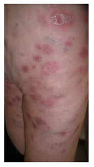
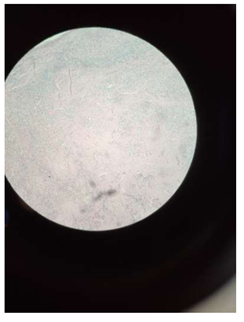
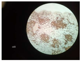
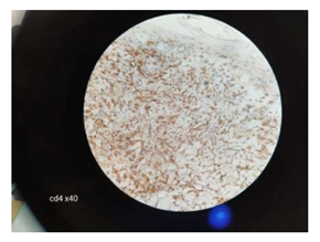
A 66-year-old female without significant past medical history presented in July 2020 to an outpatient surgery center with a two-week history of painful, rapidly growing plaques on her shins. The nodules partially regressed after systemic antibiotics (azithromycin), but new lesions appeared on the thighs and glutes. In February 2021 she presented to our dermatology clinic with many 2-5 cm, well but also ill-defined, squamous, red-violaceous, regular and irregular infiltrated plaques on her trunk and her extremities (Figure 1, 2). Some lesions were indurated and tender to palpation, whereas others had small areas of excoriation with crusting. She had malaise, weight loss and night sweats. A skin biopsy from the right arm revealed numerous small and medium sized (Figure 3) monomorphous CD2+, CD3+ (Figure 4), CD4+ (Figure 5), CD5+ CD7+ CD8- CD30- CD56- TCL-1- CD25- CD20- CD79a- Tdt- CD34- CD10- PD-1+ (Figure 4, 5) T-cell lymphoid infiltrates mainly in the dermis. Immunohistochemical staining for Ki67 expression revealed a 50% proliferative index. Subsequent molecular study showed clonal rearrangement of the T-cell receptor gene (TcRγ+). Blood cell count revealed anemia (Ht=32%), low white cell (WBC=1700 k/μL) and elevated levels of LDH (508 U/L) and ferritin (560 ng/ml). However, platelet count appeared within normal range (PLT: 171 103/μL). A bone marrow biopsy revealed no lymphoid infiltration. To rule out systemic involvement, a full body CT showed no evidence of enlarged lymph nodes. The patient did not survive to complete the planned course of chemotherapy. The term “Mature” T-Cell Lymphoma (MTCL) reflects that these cells have undergone TCR rearrangement in the thymus. For the most part, MTCLs are considered aggressive diseases and usually require multi-agent chemotherapy, and sometimes require stem cell transplantation.

Figure 1: Multiple 2-5 cm, well but also ill-defined, squamous, red-violaceous, regular and irregular infiltrated plaques.

Figure 2: Clinical picture of the trunk.

Figure 3: Monomorphous lymphocyte infiltration in the dermis (x10).

Figure 4: CD3+ lymphocytes (x20).

Figure 5: CD4+ lymphocytes (x40).
2. Conclusion
Phenotypically, PTCL-NOS show expressivity for most of the T-cell antigens which include CD2, CD3, CD4, CD5 and CD7. They usually originate from CD4 cells, but rare cases can be just CD8+, or both CD4 and CD8. The PD-1/PD-L1 pathway seems to be a potential prognostic biomarker for PTCL-NOS [3]. The presences of PD-1+ T cells in our case confirm the aggressive nature of the PTCL and further suggests its association with even more poor clinical outcomes. The diagnosis of T-cell lymphomas is highly challenging and requires an integrated approach in which clinical, morphologic, immunophenotypic and molecular data are employed.
Dermatopathologists experience the same difficulties as clinicians in the correct identification of CLD, so the cooperation between them is crucial for the prompt and correct diagnosis. In this complex context, immediate immunohistochemical studies and hematologists’ contribution are vital in the achievement of an early diagnosis. We attempt to raise awareness of the significance of including a rare underlying malignancy in the differential diagnosis when confronting a rapidly evolving eruption, along with the overall clinical deterioration of the patient. A timely diagnosis of an aggressive malignancy, as in this case, can affect the subsequent outcome.
Conflicts of Interest
None
Financial Support
None
References
- Steven H, Swerdlow, Elias Campo, et al. The 2016 revision of the World Health Organization classification of lymphoid neoplasms. Blood 127 (2016): 2375-2390.
- Broccoli A, Zinzani PL. Peripheral T-cell lymphoma, not otherwise specified. Blood 129 (2017): 1103-1112.
- Kim S, Kwon D, Koh J, et al. Clinicopathological features of programmed cell death-1 and programmed cell death-ligand-1 expression in the tumor cells and tumor microenvironment of angioimmunoblastic T cell lymphoma and peripheral T cell lymphoma not otherwise specified. Virchows Arch 477 (2020): 131-142.
