Deep Infiltrating Endometriosis in Dizygotic Twins: A Case Report and a Review of Literature
Article Information
Brittany Roberts1, Maha Al Jumaily1, Emad Mikhail2*
1Department of Obstetrics and Gynecology, University of South Florida, Morsani College of Medicine, Tampa, FL, USA
2Division of Gynecologic Subspecialities, Department of Obstetrics and Gynecology, University of South Florida, Morsani College of Medicine, Tampa, FL, USA
*Corresponding author: Emad Mikhail, MD. Department of Obstetrics and Gynecology, University of South Florida, 2 Tampa General Circle, Tampa, FL 336060, USA.
Received: 07 September 2022; Accepted: 14 September 2022; Published: 22 September 2022
Citation: Brittany Roberts, Maha Al Jumaily, Emad Mikhail. Deep Infiltrating Endometriosis in Dizygotic Twins: A Case Report and a Review of Literature. Journal of Women’s Health and Development 5 (2022): 230-234.
Share at FacebookAbstract
There is a large body of literature supporting the heritable nature of endometriosis. Although there is significant evidence demonstrating the heritability of endometriosis, there is little to no evidence demonstrating the role of heritability in patients with Deep Infiltrating Endometriosis (DIE).We present a case of two 26-year-old G0 dizygotic twins with almost identical severe posterior compartment DIE who both underwent fertility sparing laparoscopic excision of DIE.This case demonstrates an almost identical presentation of DIE in dizygotic twins. Both patients had disease manifestation in their mid 20s, similar symptomatology, almost identical intraoperative findings, and the same improvement in pain following the procedure. Although this case study further contributes to the large body of literature demonstrating the heritability of endometriosis, this is the first case study demonstrating a striking resemblance of rectovaginal DIE in dizygotic twins.
Keywords
Endometriosis; Deep infiltrating endometriosis; Hereditability; Twins
Endometriosis articles Endometriosis Research articles Endometriosis review articles Endometriosis PubMed articles Endometriosis PubMed Central articles Endometriosis 2023 articles Endometriosis 2024 articles Endometriosis Scopus articles Endometriosis impact factor journals Endometriosis Scopus journals Endometriosis PubMed journals Endometriosis medical journals Endometriosis free journals Endometriosis best journals Endometriosis top journals Endometriosis free medical journals Endometriosis famous journals Endometriosis Google Scholar indexed journals Deep infiltrating endometriosis articles Deep infiltrating endometriosis Research articles Deep infiltrating endometriosis review articles Deep infiltrating endometriosis PubMed articles Deep infiltrating endometriosis PubMed Central articles Deep infiltrating endometriosis 2023 articles Deep infiltrating endometriosis 2024 articles Deep infiltrating endometriosis Scopus articles Deep infiltrating endometriosis impact factor journals Deep infiltrating endometriosis Scopus journals Deep infiltrating endometriosis PubMed journals Deep infiltrating endometriosis medical journals Deep infiltrating endometriosis free journals Deep infiltrating endometriosis best journals Deep infiltrating endometriosis top journals Deep infiltrating endometriosis free medical journals Deep infiltrating endometriosis famous journals Deep infiltrating endometriosis Google Scholar indexed journals Hereditability articles Hereditability Research articles Hereditability review articles Hereditability PubMed articles Hereditability PubMed Central articles Hereditability 2023 articles Hereditability 2024 articles Hereditability Scopus articles Hereditability impact factor journals Hereditability Scopus journals Hereditability PubMed journals Hereditability medical journals Hereditability free journals Hereditability best journals Hereditability top journals Hereditability free medical journals Hereditability famous journals Hereditability Google Scholar indexed journals Twins articles Twins Research articles Twins review articles Twins PubMed articles Twins PubMed Central articles Twins 2023 articles Twins 2024 articles Twins Scopus articles Twins impact factor journals Twins Scopus journals Twins PubMed journals Twins medical journals Twins free journals Twins best journals Twins top journals Twins free medical journals Twins famous journals Twins Google Scholar indexed journals abdominal skin articles abdominal skin Research articles abdominal skin review articles abdominal skin PubMed articles abdominal skin PubMed Central articles abdominal skin 2023 articles abdominal skin 2024 articles abdominal skin Scopus articles abdominal skin impact factor journals abdominal skin Scopus journals abdominal skin PubMed journals abdominal skin medical journals abdominal skin free journals abdominal skin best journals abdominal skin top journals abdominal skin free medical journals abdominal skin famous journals abdominal skin Google Scholar indexed journals anterior abdominal wall articles anterior abdominal wall Research articles anterior abdominal wall review articles anterior abdominal wall PubMed articles anterior abdominal wall PubMed Central articles anterior abdominal wall 2023 articles anterior abdominal wall 2024 articles anterior abdominal wall Scopus articles anterior abdominal wall impact factor journals anterior abdominal wall Scopus journals anterior abdominal wall PubMed journals anterior abdominal wall medical journals anterior abdominal wall free journals anterior abdominal wall best journals anterior abdominal wall top journals anterior abdominal wall free medical journals anterior abdominal wall famous journals anterior abdominal wall Google Scholar indexed journals nerves articles nerves Research articles nerves review articles nerves PubMed articles nerves PubMed Central articles nerves 2023 articles nerves 2024 articles nerves Scopus articles nerves impact factor journals nerves Scopus journals nerves PubMed journals nerves medical journals nerves free journals nerves best journals nerves top journals nerves free medical journals nerves famous journals nerves Google Scholar indexed journals ureters articles ureters Research articles ureters review articles ureters PubMed articles ureters PubMed Central articles ureters 2023 articles ureters 2024 articles ureters Scopus articles ureters impact factor journals ureters Scopus journals ureters PubMed journals ureters medical journals ureters free journals ureters best journals ureters top journals ureters free medical journals ureters famous journals ureters Google Scholar indexed journals pleura articles pleura Research articles pleura review articles pleura PubMed articles pleura PubMed Central articles pleura 2023 articles pleura 2024 articles pleura Scopus articles pleura impact factor journals pleura Scopus journals pleura PubMed journals pleura medical journals pleura free journals pleura best journals pleura top journals pleura free medical journals pleura famous journals pleura Google Scholar indexed journals pericardium articles pericardium Research articles pericardium review articles pericardium PubMed articles pericardium PubMed Central articles pericardium 2023 articles pericardium 2024 articles pericardium Scopus articles pericardium impact factor journals pericardium Scopus journals pericardium PubMed journals pericardium medical journals pericardium free journals pericardium best journals pericardium top journals pericardium free medical journals pericardium famous journals pericardium Google Scholar indexed journals
Article Details
1. Introduction
Endometriosis is defined as the benign proliferation of endometrial glands and stroma outside of the endometrial cavity. Endometriosis has been demonstrated to affect 6-10% of women in their reproductive years and anywhere from 35-50% of women with pain or infertility [1]. Endometriosis may be subdivided into pelvic and extrapelvic endometriosis. Pelvic endometriosis may involve any pelvic peritoneal surfaces including the subperitoneal fat, rectovaginal space, or ovaries and is thought to occur mainly through retrograde menstruation. Extrapelvic endometriosis includes the bladder, bowel, deep pelvic nerves, ureters, anterior abdominal wall, abdominal skin, diaphragm, pleura, lungs, pericardium, and brain [2]. Symptoms may include painful periods, painful intercourse, and painful bowel movements. From a surgical perspective, there are three different types of endometriosis phenotypes including peritoneal disease, ovarian endometriomas, and Deep Infiltrating Endometriosis (DIE). Deep infiltrating endometriosis, now defined as adenomyosis externa, is a form of endometriosis characterized by histological and immunohistochemical differences [3]. There is a large body of literature supporting the heritable nature of endometriosis. Twin studies have estimated the heritability of endometriosis to be around 52% [4]. Although there is significant evidence demonstrating the heritability of endometriosis, there is little to no evidence demonstrating the role of heritability in patients with deep infiltrating endometriosis. If these are considered two similar, yet separate entities, there may be a different inheritance when discussing patients with DIE. This case presentation of DIE in dizygotic twins demonstrates the heritable nature of DIE.
2. Case Presentation
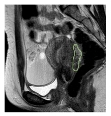
Both patients provided an informed consent, their anonymity was maintained during this case report presentation. Patient 1 was a 26-year-old G0 female who was referred for suspicion for endometriosis after being treated for suspected PID in the ED. Of note, patient was negative for gonorrhea, chlamydia, and trichomonas. She reported the onset of her symptoms 10 year prior to initial evaluation, which had increased in intensity since that time. She described the pain as occurring 2 days prior to her menses, worse on the second day, and improved by the third day. She had attempted combined oral contraceptives 6 months prior to evaluation however had little to no improvement. She described her pain as sharp, present throughout the entire abdomen, with extreme pain with bowel movements during her menstrual periods. She was not sexually active at the time of evaluation however did desire further childbearing. For workup, the patient had a transvaginal ultrasound and pelvic MRI performed. The transvaginal ultrasound demonstrated a retroverted uterus, normal in size, with heterogeneous myometrium suggestive of adenomyosis. The ovaries were noted to be fixed bilaterally to the uterus. Additionally, there was a hypoechoic nodular area seen in the posterior cul de sac measuring 20 x 8 mm. On MRI, the patient was noted to have hypointense scarring and tethering involving the posterior mid body of the uterus consistent with deep infiltrative endometriosis. Deeply infiltrative endometriosis extends roughly 4 cm in cranial caudal dimension and 4 cm in transverse dimension (see Figure 1, highlighted in green).

Figure 1: MRI demonstrating rectovaginal deep infiltrating endometriosis of patient 1.
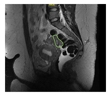
The patient was counseled on these findings including the process of deep infiltrating endometriosis and the concern for rectal scarring. The patient was recommended to have evaluation by colorectal surgery and the decision was made to proceed to the operating room for laparoscopic excision of the endometriosis. Intra operative findings included fibrosed fallopian tubes with patency proven on chromopertubation, bilateral small sized ovaries, and a large 2 x 2 cm rectovaginal nodule involving the anterior rectal wall and the posterior vaginal and both uterosacral ligaments. She underwent mobilization of the rectosigmoid, dissection of the rectovaginal septum, excision of deep infiltrating endometriosis of the rectovaginal septum, bilateral ureterolysis, and cystoscopy with attempted ureteric stenting. Final pathology was consistent with fibroadipose tissue with focal involvement by endometriosis. The patient had visits at 2 and 6 weeks post op. She was noted to have significant improvement in her pain. 5 months following the initial patient’s surgery, her twin sister presented for evaluation. She was 27 at the time of initial evaluation and was also gravida 0 para 0. The patient reported similar symptoms of her twin sister including pelvic pain which started several years prior to evaluation. She reported her pain had become significantly worse over the years and was extremely painful prior to menses and during menses. She had also attempted medical management with OCPs. She had undergone a similar work up for deep infiltrating endometriosis including a transvaginal ultrasound and pelvic MRI. Her ultrasound demonstrated a normal uterus with fixed right and left ovaries. Her left ovary was noted to have a 70 x 59 x 30 mm endometrioma present. Although the uterus was normal in appearance, it was fixed in the pelvis.

Figure 2: MRI findings of similar rectovaginal deep infiltrating endometriosis for patient 2.
On pelvic MRI (Figure 2), the uterus was noted to be normal in size and appearance. She was also noted to have a left adnexal lesion which was measured to be 6.8 x 4.2 cm. There was also noted to be focal tethering of the rectosigmoid to the left adnexal lesion however no invasion into the rectosigmoid wall. The patient was counseled on the findings and referred to colorectal surgery for a pre-operative evaluation. She then decided to proceed with surgical intervention. Intraoperative findings included deep infiltrating endometriosis of the rectovaginal septum with involvement of the anterior rectal wall and the left ureter and the left uterosacral ligament. Bilateral endometrioma were also noted with severely scarred ovaries adherent to the back of the uterus and the rectosigmoid. The patient underwent laparoscopic excision of deep infiltrating endometriosis, mobilization of the rectosigmoid, left ureterolysis, bilateral ovarian cystectomies, chromopertubation, and cystoscopy with bilateral ureteric stenting. Pathology was consistent with endometriosis involving smooth muscle and vascular adipose tissue and endometriosis involving fibromuscular connective tissues. At her post-operative visits, she was noted to have significant improvement in her pain.
3. Discussion
This case demonstrates an almost identical presentation of DIE in dizygotic twins. Both patients had disease manifestation in their mid 20s, similar symptomatology, almost identical intraoperative findings, and the same improvement in pain following the procedure. Although this case study further contributes to the large body of literature demonstrating the heritability of endometriosis, this is the first case study demonstrating a striking resemblance of rectovaginal DIE in dizygotic twins. Deep infiltrating endometriosis, also known as adenomyosis externa, is considered to be a different phenotype from ovarian endometriomas and superficial peritoneal lesions [5]. It is considered the most aggressive, severe form of the disease. The most common presentation of DIE includes the rectovaginal septum and uterosacral ligaments (69.2% of cases), the vaginal (14.5%), alimentary tract (9.9%), and urinary tract (6.4%) [6]. DIE has been demonstrated a significant role in infertility, with evidence demonstrating up to 84% success in conception following surgical resection [7]. Unlike peritoneal disease, recurrence rates of DIE after surgery are estimated to be as low as 5% [8,9]. The pathogenesis of endometriosis is multifactorial. Although a clear genetic predisposition has been identified, there are a number of contributing factors to the disease expression. These include a combination of genetics, immunological factors, and environmental factors. Endometriosis is a polygenic disease and the genes and their variants playing a role in the development of endometriosis have been widely investigated [10]. The mainstays of investigating the genetics of endometriosis include candidate gene studies, linkage and association studies, and Genome-Wide Association Studies (GWAS). To date, there are over 50 single nucleotide polymorphisms and approximately 40 endometriosis associated gene that are related to the development of endometriosis confirmed by gene association studies and GWAS [11]. Endometriosis is a complex multifactorial disease with a large body of literature supporting the heritability. We demonstrate a case of dizygotic twins with an almost identical presentation of posterior compartment deep infiltrating endometriosis including similar ultrasound/MRI findings, intraoperative findings, and post operative resolution of symptoms. To date, there have been a number of studies investigating the genetics of endometriosis with the mainstays of investigation including candidate gene studies, linkage and association studies, and Genome-Wide Association Studies (GWAS). This case provides further evidence to support the inheritance of the disease.
References
- Giudice LC, Kao LC. Lancet 364 (2004): 1789-1799.
- Bulun SE. N Engl J Med 15 (2009): 268-279.
- Liu X, Zhang Q, Guo SW.Histological and Immunohistochemical Characterization of the Similarity and Difference Between Ovarian Endometriomas and Deep Infiltrating Endometriosis. Reprod Sci 25 (2018): 329-340.
- Treloar SA, O’Connor DT, O’Connor VM, et al.Genetic influences on endometriosis in an Australian twin sample. Fertil Steril 71 (1999): 701-710.
- Tosti C, Pinzauti S, Santulli P, et al.Pathogenetic Mechanisms of Deep Infiltrating Endometriosis.Reprod Sci 22 (2015): 1053-1059.
- Clement, Philip B.MD The Pathology of Endometriosis, Advances in Anatomic Pathology 4 (2007): 241-260.
- Donnez J, Squifflet J.Complications, pregnancy and recurrence in a prospective series of 500 patients operated on by the shaving technique for deep rectovaginal endometriotic nodules. Hum Reprod 25 (2010): 1949-1958.
- Roman H, Vassilieff M, Gourcerol G, et al.:Surgical management of deep infiltrating endometriosis of the rectum: pleading for a symptom-guided approach. Hum Reprod 26 (2011): 274-281.
- De Cicco C, Corona R, Schonman R, et al.Bowel resection for deep endometriosis: a systematic review. BJOG 118 (2011): 285-291.
- Simpson JL, Elias S, Malinak LR, et al.Heritable aspects of endometriosis. I. Genetic studies. Am J Obstet Gynecol 137 (1980): 327-331.
- Vassilopoulou L, Matalliotakis M, Zervou MI, et al.:Defining the genetic profile of endometriosis. Exp Ther Med 17 (2019): 3267-3281.
