Conventional Frontal Air Sinus Imaging in Personal Identification among Adult Bangladeshi
Article Information
Farzana Maqsood1*, ShamimAra2, Md. Abdur rashid3
1Assistant Professor, Department of Anatomy, Bangladesh Medical College, Dhaka, Bangladesh
2Professor and Former Head Department of Anatomy, Dhaka Medical College, Dhaka, Bangladesh
3Professor and Head Department of Anatomy, Bangladesh Medical College, Dhaka, Bangladesh
*Corresponding Author: Farzana Maqsood, Assistant Professor, Department of Anatomy, Bangladesh Medical College, Dhaka, Bangladesh.
Received: 06 August 2023; Accepted: 10 August 2023; Published: 11 August 2023
Citation: Farzana Maqsood, ShamimAra, Md. Abdur rashid. Conventional Frontal Air Sinus Imaging in Personal Identification among Adult Bangladeshi. Journal of Analytical Techniques and Research 5 (2023): 08-12
Share at FacebookAbstract
The frontal air sinuses are essential for anatomists, radiologists, anthropologists, dentists, and clinicians to diagnose diseases. Identifying unknown persons, sex differentiation, and age determination are vital in forensic investigations because individuals' frontal air sinus characteristics make the frontal bone unique for every individual. This study aimed to determine differences in frontal air sinus width by height ratio among Bangladeshi adults. In the Department of Anatomy of Dhaka Medical College, Bangladesh, a cross-sectional analytical study was done by applying conventional and purposive sampling techniques. This study was conducted from January 2016 to December 2016. The analysis was performed on 100 radiographs of the Caldwell view of the skull of adult Bangladeshis. Out of 100 radiographs, 50 were male, and 50 were female. The Caldwell radiograph of the frontal air sinus of the selected individual was collected and viewed on the x-ray view box, traced on a transparent graph paper sheet placed on each radiograph film, and the linear measurements of frontal air sinus height and width were obtained from each radiograph by counting the boxes of transparent graph paper sheet. Then the ratio of width by size was calculated by dividing the width of the frontal air sinus by the height of the frontal air sinus individually. Unpaired Student's t-test and paired Student's t-test were done for statistical analysis of the results. The adult male left frontal air sinus width by height ratio was significantly higher than the right frontal air sinus width by height ratio, but adult female shows that right frontal air sinus width by height ratio was higher than the left frontal air sinus width by height ratio, which was not significant. Both right frontal air sinus and left frontal air sinus width by height ratio were higher in males than females, which was highly significant. The result demonstrates that the cost-effective, non-invasive, and widely applicable Caldwell view radiological morphometric analysis of frontal air sinus can be used to identify an unknown person due to the unique morphological value of the frontal air sinus in every individual.
Keywords
Frontal air sinus width by height ratio; x-ray Caldwell view
Frontal air sinus width by height ratio articles; x-ray Caldwell view articles
Frontal air sinus width by height ratio articles Frontal air sinus width by height ratio Research articles Frontal air sinus width by height ratio review articles Frontal air sinus width by height ratio PubMed articles Frontal air sinus width by height ratio PubMed Central articles Frontal air sinus width by height ratio 2023 articles Frontal air sinus width by height ratio 2024 articles Frontal air sinus width by height ratio Scopus articles Frontal air sinus width by height ratio impact factor journals Frontal air sinus width by height ratio Scopus journals Frontal air sinus width by height ratio PubMed journals Frontal air sinus width by height ratio medical journals Frontal air sinus width by height ratio free journals Frontal air sinus width by height ratio best journals Frontal air sinus width by height ratio top journals Frontal air sinus width by height ratio free medical journals Frontal air sinus width by height ratio famous journals Frontal air sinus width by height ratio Google Scholar indexed journals x-ray Caldwell view articles x-ray Caldwell view Research articles x-ray Caldwell view review articles x-ray Caldwell view PubMed articles x-ray Caldwell view PubMed Central articles x-ray Caldwell view 2023 articles x-ray Caldwell view 2024 articles x-ray Caldwell view Scopus articles x-ray Caldwell view impact factor journals x-ray Caldwell view Scopus journals x-ray Caldwell view PubMed journals x-ray Caldwell view medical journals x-ray Caldwell view free journals x-ray Caldwell view best journals x-ray Caldwell view top journals x-ray Caldwell view free medical journals x-ray Caldwell view famous journals x-ray Caldwell view Google Scholar indexed journals anatomists articles anatomists Research articles anatomists review articles anatomists PubMed articles anatomists PubMed Central articles anatomists 2023 articles anatomists 2024 articles anatomists Scopus articles anatomists impact factor journals anatomists Scopus journals anatomists PubMed journals anatomists medical journals anatomists free journals anatomists best journals anatomists top journals anatomists free medical journals anatomists famous journals anatomists Google Scholar indexed journals forensic investigations articles forensic investigations Research articles forensic investigations review articles forensic investigations PubMed articles forensic investigations PubMed Central articles forensic investigations 2023 articles forensic investigations 2024 articles forensic investigations Scopus articles forensic investigations impact factor journals forensic investigations Scopus journals forensic investigations PubMed journals forensic investigations medical journals forensic investigations free journals forensic investigations best journals forensic investigations top journals forensic investigations free medical journals forensic investigations famous journals forensic investigations Google Scholar indexed journals analysis articles analysis Research articles analysis review articles analysis PubMed articles analysis PubMed Central articles analysis 2023 articles analysis 2024 articles analysis Scopus articles analysis impact factor journals analysis Scopus journals analysis PubMed journals analysis medical journals analysis free journals analysis best journals analysis top journals analysis free medical journals analysis famous journals analysis Google Scholar indexed journals Anatomy articles Anatomy Research articles Anatomy review articles Anatomy PubMed articles Anatomy PubMed Central articles Anatomy 2023 articles Anatomy 2024 articles Anatomy Scopus articles Anatomy impact factor journals Anatomy Scopus journals Anatomy PubMed journals Anatomy medical journals Anatomy free journals Anatomy best journals Anatomy top journals Anatomy free medical journals Anatomy famous journals Anatomy Google Scholar indexed journals sampling technique articles sampling technique Research articles sampling technique review articles sampling technique PubMed articles sampling technique PubMed Central articles sampling technique 2023 articles sampling technique 2024 articles sampling technique Scopus articles sampling technique impact factor journals sampling technique Scopus journals sampling technique PubMed journals sampling technique medical journals sampling technique free journals sampling technique best journals sampling technique top journals sampling technique free medical journals sampling technique famous journals sampling technique Google Scholar indexed journals radiograph articles radiograph Research articles radiograph review articles radiograph PubMed articles radiograph PubMed Central articles radiograph 2023 articles radiograph 2024 articles radiograph Scopus articles radiograph impact factor journals radiograph Scopus journals radiograph PubMed journals radiograph medical journals radiograph free journals radiograph best journals radiograph top journals radiograph free medical journals radiograph famous journals radiograph Google Scholar indexed journals morphological cranium articles morphological cranium Research articles morphological cranium review articles morphological cranium PubMed articles morphological cranium PubMed Central articles morphological cranium 2023 articles morphological cranium 2024 articles morphological cranium Scopus articles morphological cranium impact factor journals morphological cranium Scopus journals morphological cranium PubMed journals morphological cranium medical journals morphological cranium free journals morphological cranium best journals morphological cranium top journals morphological cranium free medical journals morphological cranium famous journals morphological cranium Google Scholar indexed journals morphometric articles morphometric Research articles morphometric review articles morphometric PubMed articles morphometric PubMed Central articles morphometric 2023 articles morphometric 2024 articles morphometric Scopus articles morphometric impact factor journals morphometric Scopus journals morphometric PubMed journals morphometric medical journals morphometric free journals morphometric best journals morphometric top journals morphometric free medical journals morphometric famous journals morphometric Google Scholar indexed journals
Article Details
Introduction:
The frontal air sinuses are paired, irregularly shaped, air-conditioning cavity lined by mucoperiosteum and are located posterior to the superciliary arches and at the roof of the nose between the outer and inner tables of the frontal bone [1]. The frontal air sinus divided into two chambers, right and left frontal air sinus. In between right and left frontal air sinus lies a septum which usually deviates from the midline. The right and left frontal air sinus chambers are rarely symmetrical [2]. Due to the independent development of each sinus, the right and left frontal air sinus chambers are typically asymmetrical. The sinuses are subdivided into several recesses and through incomplete bony septa the recesses are communicate with each other [3]. The frontal air sinus is a triangular, pyramid-shaped cavity extending between the two tables of the ascending portion of the frontal bone [4]. The frontal air sinus and the nasal fossa communicates through ethmoidal infundibulum. It begins to develop during the second year of life, reaches its maximum size at the age of twenty, and remains stable through the rest of life [5]. The frontal sinus development shows a growth rhythm similar to body height development with a well-defined pubertal peak. The frontal air sinus embryonically develops from an ethmoidal cell which is not visible at birth [6-7]. Even though during birth the frontal air sinus bud is present in the ethmoid region, it is actually radiologically evident after six years when it projects above the orbital rim [6]. Pneumatization of the skull is strictly connected with the morphology and degree of development of paranasal sinuses [8]. The frontal air sinus area, size, and shape vary from one person to another person and one race to another race [9-10]. The frontal air sinus usually shows unilateral or hypoplasia but Complete aplasia is very rare[11]. The hardness of the frontal air sinus may lead to a misbalance in the human cranium [12]. Frontal air sinus pneumatization depends on individuals growth differences and mucosal desorption processes, the frontal bone quality which is to be pneumatized, the pressure exerted by the growing brain on the internal lamina of the frontal air sinus, the various pressure and hydrodynamic conditions of the cranium that influences the blood supply of the frontal sinus area and hereditary factor, Genetic diseases, environmental condition, and past infection also affects the pneumatization process of the frontal air sinus within the frontal bone [8-9]. Pneumatization of the skull is strictly connected with the morphology and degree of development of paranasal sinuses [13]. The plain radiographic study provides knowledge of the anatomy, size, capacity, asymmetry, and variants of the frontal air sinus for specific populations. That knowledge is necessary for the clinician in pathologies of the acute and chronic sinuses and surgical procedures [14]. There is a correlation between frontal sinusitis and the possible complications from the orbit when the frontal air sinuses unusually extends above the orbital roof and these cases are considered high-risk cases [15]. The frontal air sinus study helps surgeons to avoid or minimize possible complications by providing clear knowledge about the variations that may predispose patients to increased risk of intraoperative complications [16].The irregular shape and distinctive characteristics of the frontal air sinus vary from person to person and make the frontal bone unique for every individual Yoshino [10]. Even in monozygotic twins, the frontal air sinus plays a similar role in fingerprints and DNA tests as each frontal air sinus shows unique morphological value, and this distinctive characteristic of the frontal air sinus helps in the forensic identification of unknown deceased persons [17-18]. The frontal sinus is also smaller in females than in males because of morphological cranium differences between males and females [19]. The morphometric knowledge of sexual dimorphism also plays a vital role in the determination of the gender of an unknown person in forensic investigation, especially in cases where only fragments of the skull remain and there is no possibility of identification [20]. To confirm the identification of highly decomposed or burnt or disfigured human remains, radiographic comparison of osteologic structures like frontal air sinus has been commonly used [20]. Radiological identification of frontal air sinuses has a very important place in forensic medicine, particularly in the lack of comparative evidence like DNA samples and fingerprints [21]. The human skull has been massively studied and involves the comparison of antemortem radiographs, usually carried out for clinical reasons, with post-mortem radiographs taken solely for the identification of specific, individualizing structures. As the chance of two people having the same morphology of frontal air sinus is extremely rare, the radiographic comparison of frontal air sinus dimension is a scientifically secure method for the identification of unknown dead bodies [22-23]. The study was done to evaluate and compare the width by height ratio of the frontal air sinus on a Caldwell view radiograph and to use the data to determine and establish the identity of an individual.
Materials & Methods:
The cross-sectional analytical study was carried out by applying a convenient and purposive sampling technique in the Department of Anatomy, Dhaka Medical College, Bangladesh, from January 2016 to December 2016. The study was performed on 100 radiographs of Caldwell's view of the skull of adult Bangladeshis. Out of 100 radiographs, 50 were male and 50 were female, with ages ranging from 20 to 50 years. Individuals with a history or X-ray showing any feature of sinusitis, repeated common cold, chronic headache, trauma or surgery of the skull or face including eye, ear, and nose, and clinical features of endocrine disturbances or systemic disorder or any sort of cranial asymmetry or facial asymmetry were excluded from the study. The X-ray machine was used for doing skull radiographs in Caldwell view. The study subject was in an erect posture in the posterior - anterior position in front of a part of the x-ray machine facing stand greed. The cassette of the stand greed machine had an imaging plate that was kept towards the head position. The vertical axis was parallel to the film in Caldwell projection and no space between the frontal bone and the cassette. The center of the forehead and nose was adjusted in a position so that the midsagittal plane was perpendicular to the plane of the film, and the orbitomeatal line formed an angle of 15 degrees from the plane of the film. Then the head was immobilized in that position, and the shoulder was adjusted in the same horizontal plane. The subject was advised to suspend respiration for the exposure. Using a well-collimated beam, the horizontal central ray is centered in the midline to the occipital region at the level of the lower orbital margin. Radiation was projected on the imaging plate, and it was transformed in the monitor for editing and zooming of the radiograph. It was zoomed at 100%. AGFA printer was used for printing the X-ray film. The radiographs were collected from every selected individual and viewed on the x-ray view box and transparent graph paper sheet was placed on the view box containing the radiograph, and the frontal air sinus was traced on a transparent graph paper sheet placed on each radiograph film, and then the different dimensions of the frontal air sinus was measured. All the measurement of the frontal sinus was taken by drawing a tangential line above the superior border of the two orbits because the inferior limit of the frontal air sinus is not so clear [10].
The measurement was done according to the sequence below by Camargo et al. [24].
-At first, the radiography was placed on a view box, and the transparent graph paper sheet was placed on the radiograph.
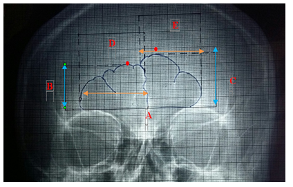
-The baseline (A) was drawn directly on the transparent graph paper at the level of the superior border or upper limit of two orbits (inferior border of the frontal sinus), as illustrated in the figure.
-The separation between the right and left frontal sinus was based on the frontal inter-sinus septum in order to permit quantifying one width on each side.
-The height of each side (B and C) was determined by marking and drawing a straight line between the baseline and upper limit of the frontal sinus.
-The largest width (D and E) of the frontal sinus was determined by marking and drawing straight lines from the maximum distance between the medial and lateral lines of the right and left side of the frontal sinus.
-Linear measurements of height and width of the frontal air sinus were obtained from each radiograph by counting the boxes of graph paper; height and width were expressed in centimeters (cm).
-Square box was counted within the sinus, and an area covering more than 50% of the square was considered.
-All these measurements were obtained by the portion projecting above the baseline. The separation of the right and left sides of the frontal sinus was based on the inter-sinus septum, which denotes the margin between the two main sinus cavities.
-The ratio of the width by height of the frontal air sinus was calculated by dividing the width of the frontal air sinus by the height of the frontal air sinus individually [25].
Paired Student t-tests were done to compare the differences between the height of the right and left frontal air sinus and the width of the right and left frontal air sinus. Unpaired Student's t-tests were done to analyze the differences between the right and left frontal air sinus dimensions between males and females. Statistical significance was accepted at (p<0.05).
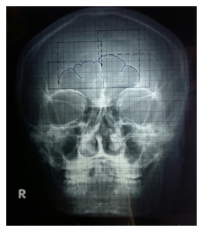
Procedure measurement from a radiograph of frontal air sinus height and width is shown in Figures 1, 2 (i), 2 (ii)

Figure 1: procedure of taking caldwell view radiograph of skull

Figure 2 (i): procedure of tracing caldwell view radiograph.

Figure 2 (ii): procedure of estimating of height, width of right and left frontal sinus.
Red dot indicate highest point of right and left frontal air sinus.
Arrow indicate height of right (line B) and left (line C)frontal air sinus
Green dot indicate widest point of right and left frontal air sinus.
Arrow indicate width of the right (line D) and left (line E) frontal air sinus.
|
Group |
Ratio of width by height of the frontal air sinus |
||
|
Right (Mean±SD) |
Left(Mean±SD) |
P value |
|
|
Male (n=50) |
0.51 ± 0.15 |
0.53 ± 0.16 |
0.114ns |
|
Female (n=50) |
0.42 ± 0.17 |
0.42±0.14 |
0.670ns |
|
P value |
0.004* |
0.001* |
|
Figures in parentheses indicate range. SD= Standard Deviation
Comparison between male and female was done by Unpaired Student’s ‘t’ test. Comparison between values of right and left frontal sinus of same group was done by Paired Student’s ‘t’ test ns= not significant, *= significant
Table1: Comparison of the Ratio between width by height of the frontal air sinus in male and female

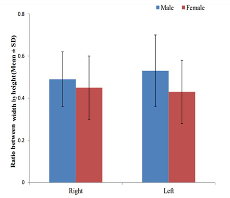
Figure 3: Comparison between ratio of width by height of right and left frontal air sinus in male and female
Ethical Clearance:
The study was approved by the ethical review committee of Dhaka Medical College, Dhaka, Bangladesh.
Result:
In males, width by height ratio of the right frontal air sinus ranged from 1.48 to 4.5 cm, the mean width by height ratio was 0.51 ± 0.15 cm and width by height ratio of the left frontal air sinus ranged from 0.89 to 5.3 cm, and the mean width by height ratio was 0.53 ± 0.16 cm. No significant difference (P=0.114) was observed in the width-by-height ratio of right and left frontal air sinuses in males. In females, width by height ratio of the right frontal air sinus ranged from 0.33 to 4.14 cm, the mean width by height ratio was 0.42 ± 0.17 cm and width by height ratio of the left frontal air sinus ranged from 0.26 to 3.2 cm and the mean width by height ratio was 0.42 ± 0.14 cm. No significant difference (P=0.670) was observed in the width by height ratio of right and left frontal air sinuses in females. The mean of right frontal air sinus width by height was observed to be significantly higher (p=0.004) in males than females. The mean of left frontal air sinus width by height ratio was observed to be significantly higher (p=0.001) in males than females.
Discussion
In the present study, the adult male left frontal air sinus width by height ratio was higher than the right frontal air sinus width by height ratio, but the adult female shows right frontal air sinus width/ height ratio was higher than the left frontal air sinus width by height ratio. Both right frontal air sinus and left frontal air sinus width by height ratio were higher in males than females. According to researcher Verma, P., et al. (2015), the adult male and female frontal air sinus width by height ratio had an average of 1.67 ± 0.04 cm and 1.62 ± 0.04 cm, respectively. The parameter shows that frontal air sinus width by height ratio was significantly higher (p<0.000) in males than in females. So, the finding of the present study is similar to the finding reported by the researcher Verma, P., et al.(2015). It was observed that the value of the study conducted by Verma was significantly higher (p>0.000) compared to the present study.
Conclusion:
The result of the present study demonstrates that the adult male left frontal air sinus width by height ratio was higher than the right frontal air sinus width by height ratio, and the adult female shows right frontal air sinus width by height ratio was higher than the left frontal air sinus width by height ratio. Both right frontal air sinus and left frontal air sinus width by height ratio were higher in males than females. This study not only plays an important role in the medico-legal aspects of forensic science, like personal identification, age determination, and gender differentiation but also helps as a guideline for anatomists, clinicians, dentists, and surgeons.
References
- Bullen, R. et al. The frontal sinus on facial plain radiographs. Journal of morphological science 27 (2010): 77-81.
- Belaldavar, C., et al. Assessment of frontal sinus dimension to determine sexual dimorphism among indian adults. Journal of Forensic Dental Sciences 6 (2014): 1-8.
- Ponde, M. J., et al. Anatomical Variation of Frontal Sinus. International Journal of Morphology 26 (2008): 803-808.
- Tiwari P. et al. The frontal sinus on facial plain radiographs. Journal of Oral Medicine, Oral Surgery, Oral Pathology and Oral Radiology 2 (2016): 11-22.
- Selarka, B. Conventional frontal sinus imaging- A tool in gender determination: original study. Journal of Oral Medicine and Radiology 5 (2015): 33-37.
- Mathur H, et al. Conventional Frontal Sinus Imaging In Identification Of Sex Original Study In Population Of Udaypur City, India. Journal of Medical Science and Clinical Research 1 (2013): 33-37.
- Al-Sheakli I I, Mohammed S A, Taha S S. The frontal sinus dimensions in mouth and nasal breathers in Iraqi adult subjests. Journal of Baghdad College Dentistry 25 (2013): 155-163.
- Silva RFD. et al. Identification of charred body through the radiographic morphology of the frontal sinus. Annals of forensic Research and Analysis 2 (2014): 18-23.
- Karakas S, Kavakli A. Morphological examination of the paranasal sinuses and mastoid air cells using computed tomography. Ann Saudi Med 25 (2005): 41-45
- Yoshino M, et al. Classification system of frontal sinus patterns by radiography. Its application to identification of unknown skeletal remains. Forensic Science International, 34 (1987): 289-299.
- Vidya C S, Shamasundar N M. Computer tomographic study of frontal sinus patterens in skulls of south Indian population. International journal of scintific and research publications 4 (2014): 2250-3153.
- Kanat A, et al. Frontal sinus asymmetry: Is it an effect of cranial asymmetry? x-ray analysis of 496 normal adult human frontal sinus. Journal of Neurosciences in Rural Practice 6 (2015): 511-514.
- Teul I, et al. Morphological Evaluation of the paranasal sinuses of the skull from different historic periods. Durham Anthropology Journal 12 (2005): 1-4.
- Ruiz R C, Wafae N. Anatomo-radiological and morphometrical study of the frontal sinus in humans. Brazilian Journal of morphological Sciences 21 (2004): 53-56.
- Natsis K. and et al. The Frontal sinus anatomical variations with potential consequences for orbit. Study on cadavers. Morphologie 88 (2004): 35-38.
- Patil A A, Revankar A V. Reliability of the frontal sinus index as a maturity indicator. India. Indian Journal of Dental Research. 24 (2013): 523.
- Neha, et al. Morphometric evaluation of frontal sinus in relation to gender-a forensic study. Journal of Dental Science 1 (2015): 7-11.
- Nambiar P, Naidu M D K, Subramaniam K. Anatomical variability of the frontal sinuses and their application in forensic identification Clin Anat 12 (1999): 16–19.
- Soman B A, Sujata G P Lingappa A. Morphometric evaluation of frontal sinus in relation to age and gender in subjects residing in Davagere, Karnataka. Journal of forensic Dental Sciences 8 (2016): 1-8.
- Verma P, et al. Combined use of frontal sinus and nasal septum patterns as an aid in forensics: A digital radiographic study. North American Journal Of Medical Sciences 7 (2015): 47-52.
- Nikam S S, et al. Personal Identification in Forensic Science Using Uniqueness of Radiogragic Image of Frontal Sinus. Journal of forensic odontosomatology 33 (2015): 1-7.
- Soares C B R B S, et al. Human identification study by means of frontal sinus imaginological aspects. Forensic science international 262 (2016): 183-189.
- Bengiac A G, Thiel B A, Haba D. Reliability of the frontal sinus index for sex determination using CBCT. Romanian society of legal medicine 6(2015): 275-278.
- Camargo R. et al. The frontal sinus morphology in radiograph of Brazilian subjects: its forensic importance. Brazilian journal of morphological science 24 (2007): 239-243.
- Verma S, Mahima V G, Patil K. Radio morphometric analysis of frontal air sinus for sex determination. Journal of forensic DentalSciences6(2014):177-182.
