Congenital Atrichia with Recurrent Episcleritis-A Rare Case Report
Article Information
Anubhav Chauhan1*, Shveta Chauhan2, Mandeep Tomar1
1Department of Ophthalmology, Dr Yashwant Singh Parmar Govt. Medical College, Nahan, District Sirmour, Himachal Pradesh, India
2Pine Castle, Near Mist Chamber, Khalini, Shimla, Himachal Pradesh, India
*Corresponding Author: Dr. Anubhav Chauhan, Department of Ophthalmology, Dr Yashwant Singh Parmar Govt. Medical College, Nahan, District Sirmour, Himachal Pradesh
Received: 10 May 2019; Accepted: 20 May 2019; Published: 28 May 2019
Citation: Anubhav Chauhan, Shveta Chauhan, Mandeep Tomar. Congenital Atrichia with Recurrent Episcleritis-A Rare Case Report. Archives of Nephrology and Urology 2 (2019): 026-028.
Share at FacebookAbstract
Congenital atrichia is a rare disorder and is often associated with various systemic diseases. Ocular involvement has also been reported. We report a rare case of congenital atrichia with recurrent episcleritis. To the best of our knowledge and after an extensive internet search, this is probably the first reported case of congenital atrichia with episcleritis. The association may be a co-incidental finding or may have a broader perspective. Hence, further studies are warranted.
Keywords
Renal, Nephrotic syndrome, Keratitis, Congenital nystagmus, Cone-rod dysfunction
Renal articles, Nephrotic syndrome articles, Keratitis articles, Congenital nystagmus articles, Cone-rod dysfunction articles
Renal articles Renal Research articles Renal review articles Renal PubMed articles Renal PubMed Central articles Renal 2023 articles Renal 2024 articles Renal Scopus articles Renal impact factor journals Renal Scopus journals Renal PubMed journals Renal medical journals Renal free journals Renal best journals Renal top journals Renal free medical journals Renal famous journals Renal Google Scholar indexed journals Nephrotic syndrome articles Nephrotic syndrome Research articles Nephrotic syndrome review articles Nephrotic syndrome PubMed articles Nephrotic syndrome PubMed Central articles Nephrotic syndrome 2023 articles Nephrotic syndrome 2024 articles Nephrotic syndrome Scopus articles Nephrotic syndrome impact factor journals Nephrotic syndrome Scopus journals Nephrotic syndrome PubMed journals Nephrotic syndrome medical journals Nephrotic syndrome free journals Nephrotic syndrome best journals Nephrotic syndrome top journals Nephrotic syndrome free medical journals Nephrotic syndrome famous journals Nephrotic syndrome Google Scholar indexed journals Keratitis articles Keratitis Research articles Keratitis review articles Keratitis PubMed articles Keratitis PubMed Central articles Keratitis 2023 articles Keratitis 2024 articles Keratitis Scopus articles Keratitis impact factor journals Keratitis Scopus journals Keratitis PubMed journals Keratitis medical journals Keratitis free journals Keratitis best journals Keratitis top journals Keratitis free medical journals Keratitis famous journals Keratitis Google Scholar indexed journals Congenital Atrichia articles Congenital Atrichia Research articles Congenital Atrichia review articles Congenital Atrichia PubMed articles Congenital Atrichia PubMed Central articles Congenital Atrichia 2023 articles Congenital Atrichia 2024 articles Congenital Atrichia Scopus articles Congenital Atrichia impact factor journals Congenital Atrichia Scopus journals Congenital Atrichia PubMed journals Congenital Atrichia medical journals Congenital Atrichia free journals Congenital Atrichia best journals Congenital Atrichia top journals Congenital Atrichia free medical journals Congenital Atrichia famous journals Congenital Atrichia Google Scholar indexed journals Congenital nystagmus articles Congenital nystagmus Research articles Congenital nystagmus review articles Congenital nystagmus PubMed articles Congenital nystagmus PubMed Central articles Congenital nystagmus 2023 articles Congenital nystagmus 2024 articles Congenital nystagmus Scopus articles Congenital nystagmus impact factor journals Congenital nystagmus Scopus journals Congenital nystagmus PubMed journals Congenital nystagmus medical journals Congenital nystagmus free journals Congenital nystagmus best journals Congenital nystagmus top journals Congenital nystagmus free medical journals Congenital nystagmus famous journals Congenital nystagmus Google Scholar indexed journals Cone-rod dysfunction articles Cone-rod dysfunction Research articles Cone-rod dysfunction review articles Cone-rod dysfunction PubMed articles Cone-rod dysfunction PubMed Central articles Cone-rod dysfunction 2023 articles Cone-rod dysfunction 2024 articles Cone-rod dysfunction Scopus articles Cone-rod dysfunction impact factor journals Cone-rod dysfunction Scopus journals Cone-rod dysfunction PubMed journals Cone-rod dysfunction medical journals Cone-rod dysfunction free journals Cone-rod dysfunction best journals Cone-rod dysfunction top journals Cone-rod dysfunction free medical journals Cone-rod dysfunction famous journals Cone-rod dysfunction Google Scholar indexed journals diagnosis articles diagnosis Research articles diagnosis review articles diagnosis PubMed articles diagnosis PubMed Central articles diagnosis 2023 articles diagnosis 2024 articles diagnosis Scopus articles diagnosis impact factor journals diagnosis Scopus journals diagnosis PubMed journals diagnosis medical journals diagnosis free journals diagnosis best journals diagnosis top journals diagnosis free medical journals diagnosis famous journals diagnosis Google Scholar indexed journals non steroidal anti-inflammatory drugs articles non steroidal anti-inflammatory drugs Research articles non steroidal anti-inflammatory drugs review articles non steroidal anti-inflammatory drugs PubMed articles non steroidal anti-inflammatory drugs PubMed Central articles non steroidal anti-inflammatory drugs 2023 articles non steroidal anti-inflammatory drugs 2024 articles non steroidal anti-inflammatory drugs Scopus articles non steroidal anti-inflammatory drugs impact factor journals non steroidal anti-inflammatory drugs Scopus journals non steroidal anti-inflammatory drugs PubMed journals non steroidal anti-inflammatory drugs medical journals non steroidal anti-inflammatory drugs free journals non steroidal anti-inflammatory drugs best journals non steroidal anti-inflammatory drugs top journals non steroidal anti-inflammatory drugs free medical journals non steroidal anti-inflammatory drugs famous journals non steroidal anti-inflammatory drugs Google Scholar indexed journals
Article Details
1. Case Study
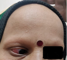
A 35-year-old female patient presented to the department of ophthalmology with a history of redness, lacrimation and photophobia in the right eye off and on for the past one year (Figure 1). There was no other ocular complaint. There was no other significant medical, surgical, family, traumatic or drug abuse history. Ocular examination was carried out and her visual acuity was 6/6 in both the eyes; pupillary reactions, ocular movements, colour vision, fundus and intraocular pressure were normal bilaterally. Slit lamp and torch light examination revealed simple episcleritis in the right eye.
There were absence of eyebrows and eyelashes on examination. On further questioning the patient, she removed her cap to reveal the total absence of hairs on the scalp. She further stated that there was a complete absence of hairs since birth and dermatological consultation had revealed congenital atrichia. Rest of her general physical and systemic examination was normal. For episcleritis, the patient was started on tablet naproxen and fluorometholone eye drops. Blood investigations to rule out connective tissue disorders were within normal levels. To the best of our knowledge and after an extensive internet search, this is probably the first reported case of association between episcleritis and congenital atrichia.

Figure 1: Patient with a history of redness, lacrimation and photophobia in the right eye.
2. Discussion
Congenital atrichia is a rare disease causing irreversible alopecia which is inherited autosomal recessively. There is complete irreversible hair loss seen soon after birth. Situs inversus and mesocardia has also been reported to be associated with this disease. Other associations include Moynahan’s syndrome (characterized by seizures with mental retardation), hidrotic ectodermal dysplasia (palmoplantar keratoderma, thickened nails) and aging syndromes [1]. Loss of scalp hair is seen between one to six months of age, after which no growth occurs. There may also be sparse or absent hairs on the eyebrow, eyelash, and body. Differential diagnosis include vitamin D dependant rickets, congenital alopecia universalis, and ectodermal dysplasia [2]. IFAP (Ichthyosis follicularis with alopecia and photophobia) syndrome has also been reported in literature where patient presents with a triad of striking alopecia, photophobia and generalized cutaneous ’thorn-like’ projections [3].
Atrichia has also been reported to be associated with anomalies of the face, nails, cartilage, speech, respiratory tract, digits, and oral cavity. Renal involvement in the form of nephrotic syndrome and glucosuria has also been reported. Ocular involvement in the form of congenital cataracts, microphtalmia, keratitis, congenital nystagmus, cone-rod dysfunction, and high myopia has been reported [4]. Episcleritis is the inflammation of the episclera (a thin, loose, highly vascular connective tissue layer between the conjunctive and sclera). It can be idiopathic, or it can be associated with connective tissue disorders like rheumatoid arthritis, scleroderma and systemic lupus erythematosus. Watson and Hayreh classified episcleritis into simple and nodular. Most patients have intermittent bouts of moderate or severe inflammation at intervals of 1–3 months. Symptoms include acute onset of redness, lacrimation, and photophobia. It commonly affects a single quadrant in one eye. Bilateral involvement suggests underlying systemic disease. Systemic non steroidal anti-inflammatory drugs (NSAIDs) and topical steroids are used in its treatment [5].
Conflicts of Interest
The authors declare that they have no competing interests.
Financial Disclosure
The authors have no proprietary or commercial interest in any material discussed in this article.
References
- Verma R, Vasudevan B, Pragasam V, et al. Congenital atrichia with papular lesions. Indian J Paediatr Dermatol 15 (2014): 94-95.
- Chouhan C, Khullar R, Rao P, et al. Atrichia congenita. Indian Dermatol Online J 6 (2015): 352-353.
- Rai VM, Shenoi SD. Ichthyosis follicularis with alopecia and photophobia (IFAP) syndrome. Indian J Dermatol Venereol Leprol 72 (2006): 136-138.
- Bennàssar A, Ferrando J, Grimalt R. Congenital atrichia and hypotrichosis.World J Pediatr 7 (2011): 111-117.
- Salama A, Elsheikh A, Alweis R. Is this a worrisome red eye? Episcleritis in the primary care setting. Journal of community hospital internal medicine perspectives 8 (2018): 46-48.
