Clinical Significance of Ultrasonography Markers in Prenatal Diagnosis of Turner Syndrome in Fetuses-90 Cases Reports
Article Information
Allybocus Zubair Akbar*
Center of Genetics and Prenatal Diagnosis, Department of Gynecology, The First affiliated hospital of Zhengzhou University, Zhengzhou, Henan province, China
*Corresponding Author: Allybocus Zubair Akbar, Center of Genetics and Prenatal Diagnosis, Department of Gynecology, The First affiliated hospital of Zhengzhou University, Zhengzhou, Henan province, China
Received: 05 February 2019; Accepted: 13 February 2019; Published: 15 February 2019
Citation: Allybocus Zubair Akbar. Clinical Significance of Ultrasonography Markers in Prenatal Diagnosis of Turner Syndrome in Fetuses-90 Cases Reports. Journal of Radiology and Clinical Imaging 2 (2019): 007-015.
Share at FacebookAbstract
Objective: To evaluate the clinical significance of ultrasonography markers in prenatal diagnosis of Turner syndrome in fetuses.
Methods: Retrospectively 90 karyotypically documented fetuses with Turner syndrome reviewed from our database between August 2010 and April 2018 made up our study population. Women were aged between 19 to 40 years and weeks of gestation from 9 to 32 weeks. Ultrasound findings documented by skilled sonographer, performed in a systemic order from fetal head to toe, indicative of turner syndrome were noted, grouped and tabulated. Advanced maternal age women had routine fetal karyotyping while for younger women, it was indicated after abnormal ultrasonic findings. Amniocentesis or chorionic villus sampling were source for fetal chromosome extraction for karyotyping. Karyotyping (G-banding) was performed under standard protocol and karyotypes were described according to the International System for Human Cytogenetic Nomenclature. The karyotypes results were grouped as pure monosomy, 45X, and Mosaics In the evaluation of clinical significance of ultrasound markers, P values lower than 0.001 were considered as statistically significant. The calculations were performed using a statistical package program.
Results: 61 fetuses were diagnosed in <30 yr group of which 48 (78%) 45X vs 17 (18%) mosaic, 17 fetuses in 30-35 yr with 7 (41%) 45X vs 10 (58.8%) mosaic and 12 fetuses in >35yr group as 6 (50%) 45X vs 6 (50%) mosaic. 32 fetuses were diagnosed as Turner syndrome in <12 weeks period of which 31 (96%) 45X vs 1 (4%) mosaic, 27 fetuses in 12-20 weeks period of which 20 (33%) 45X vs 7 (24%) and 31 in >20 weeks period as 10 (16%) 45X vs 21 (72%) mosaic. From ultrasound findings, 25 fetuses were diagnosed as intrauterine death, of which 24 (96%) 45X vs 1(4%) mosaic, (p<0.001) and 20 (80%) of them occurring in <12 weeks period (p<
Keywords
Turner syndrome, Ultrasound markers, Karyotyping, Cystic hygroma, Pleural effusion, Skin edema, Intrauterine demise
Turner syndrome, Ultrasound markers, Karyotyping, Cystic hygroma, Pleural effusion, Skin edema, Intrauterine demise
Turner syndrome articles Turner syndrome Research articles Turner syndrome review articles Turner syndrome PubMed articles Turner syndrome PubMed Central articles Turner syndrome 2023 articles Turner syndrome 2024 articles Turner syndrome Scopus articles Turner syndrome impact factor journals Turner syndrome Scopus journals Turner syndrome PubMed journals Turner syndrome medical journals Turner syndrome free journals Turner syndrome best journals Turner syndrome top journals Turner syndrome free medical journals Turner syndrome famous journals Turner syndrome Google Scholar indexed journals Ultrasound markers articles Ultrasound markers Research articles Ultrasound markers review articles Ultrasound markers PubMed articles Ultrasound markers PubMed Central articles Ultrasound markers 2023 articles Ultrasound markers 2024 articles Ultrasound markers Scopus articles Ultrasound markers impact factor journals Ultrasound markers Scopus journals Ultrasound markers PubMed journals Ultrasound markers medical journals Ultrasound markers free journals Ultrasound markers best journals Ultrasound markers top journals Ultrasound markers free medical journals Ultrasound markers famous journals Ultrasound markers Google Scholar indexed journals Karyotyping articles Karyotyping Research articles Karyotyping review articles Karyotyping PubMed articles Karyotyping PubMed Central articles Karyotyping 2023 articles Karyotyping 2024 articles Karyotyping Scopus articles Karyotyping impact factor journals Karyotyping Scopus journals Karyotyping PubMed journals Karyotyping medical journals Karyotyping free journals Karyotyping best journals Karyotyping top journals Karyotyping free medical journals Karyotyping famous journals Karyotyping Google Scholar indexed journals Cystic hygroma articles Cystic hygroma Research articles Cystic hygroma review articles Cystic hygroma PubMed articles Cystic hygroma PubMed Central articles Cystic hygroma 2023 articles Cystic hygroma 2024 articles Cystic hygroma Scopus articles Cystic hygroma impact factor journals Cystic hygroma Scopus journals Cystic hygroma PubMed journals Cystic hygroma medical journals Cystic hygroma free journals Cystic hygroma best journals Cystic hygroma top journals Cystic hygroma free medical journals Cystic hygroma famous journals Cystic hygroma Google Scholar indexed journals Pleural effusion articles Pleural effusion Research articles Pleural effusion review articles Pleural effusion PubMed articles Pleural effusion PubMed Central articles Pleural effusion 2023 articles Pleural effusion 2024 articles Pleural effusion Scopus articles Pleural effusion impact factor journals Pleural effusion Scopus journals Pleural effusion PubMed journals Pleural effusion medical journals Pleural effusion free journals Pleural effusion best journals Pleural effusion top journals Pleural effusion free medical journals Pleural effusion famous journals Pleural effusion Google Scholar indexed journals Skin edema articles Skin edema Research articles Skin edema review articles Skin edema PubMed articles Skin edema PubMed Central articles Skin edema 2023 articles Skin edema 2024 articles Skin edema Scopus articles Skin edema impact factor journals Skin edema Scopus journals Skin edema PubMed journals Skin edema medical journals Skin edema free journals Skin edema best journals Skin edema top journals Skin edema free medical journals Skin edema famous journals Skin edema Google Scholar indexed journals Intrauterine demise articles Intrauterine demise Research articles Intrauterine demise review articles Intrauterine demise PubMed articles Intrauterine demise PubMed Central articles Intrauterine demise 2023 articles Intrauterine demise 2024 articles Intrauterine demise Scopus articles Intrauterine demise impact factor journals Intrauterine demise Scopus journals Intrauterine demise PubMed journals Intrauterine demise medical journals Intrauterine demise free journals Intrauterine demise best journals Intrauterine demise top journals Intrauterine demise free medical journals Intrauterine demise famous journals Intrauterine demise Google Scholar indexed journals ultrasonography articles ultrasonography Research articles ultrasonography review articles ultrasonography PubMed articles ultrasonography PubMed Central articles ultrasonography 2023 articles ultrasonography 2024 articles ultrasonography Scopus articles ultrasonography impact factor journals ultrasonography Scopus journals ultrasonography PubMed journals ultrasonography medical journals ultrasonography free journals ultrasonography best journals ultrasonography top journals ultrasonography free medical journals ultrasonography famous journals ultrasonography Google Scholar indexed journals monosomy articles monosomy Research articles monosomy review articles monosomy PubMed articles monosomy PubMed Central articles monosomy 2023 articles monosomy 2024 articles monosomy Scopus articles monosomy impact factor journals monosomy Scopus journals monosomy PubMed journals monosomy medical journals monosomy free journals monosomy best journals monosomy top journals monosomy free medical journals monosomy famous journals monosomy Google Scholar indexed journals amniocentesis articles amniocentesis Research articles amniocentesis review articles amniocentesis PubMed articles amniocentesis PubMed Central articles amniocentesis 2023 articles amniocentesis 2024 articles amniocentesis Scopus articles amniocentesis impact factor journals amniocentesis Scopus journals amniocentesis PubMed journals amniocentesis medical journals amniocentesis free journals amniocentesis best journals amniocentesis top journals amniocentesis free medical journals amniocentesis famous journals amniocentesis Google Scholar indexed journals
Article Details
1. Introduction
Prenatal screening or diagnosis is a fundamental part of antenatal care as it enables identification of mild to severe abnormalities in the fetus. This permits proper planning and management of current and future pregnancy. Chromosomal abnormalities can either be chromosome structural defects (disjunction) or chromosome numerical anomalies (aneuploidy). Turner syndrome (TS), a rare chromosomal disorder occurring in 25-50 per 100 000 births in female, is characterized by the complete (Classic) or partial (Mosaic) absence of the second sex chromosome and associated with one or more clinical manifestations, from prenatal, postnatal to adult period, that are key for recognizing TS according to latest International guidelines [1, 2]. Due to its lower penetrance compare to the 45X monosomy [3], mosaic TS will produce phenotypes ranging from asymptomatic to symptomatic depending on number of normal cell in the body of fetus [3]. With the advent of improved ultrasound imaging, it is now possible to make a sonography diagnosis of TS. Classic sonographic findings of TS include increased nuchal translucency, diffuse fetal edema, cystic hygroma with septations, renal and cardiac anomalies and other constellation of findings postnatal [1, 2]. The key to an accurate antenatal diagnosis is careful scanning of the fetus and knowledge of the abnormalities by sonographer. Are these ultrasound findings reliable and specific to diagnose TS in fetus, we carry out a retrospective study at Zhengzhou University. While karyotyping is the gold standard, we shall see where sonography stands for diagnosis of TS in fetus.
2. Case Report
2.1 Patients
A retrospective search of our genetic and prenatal diagnosis center database between August 2010 and April 2018 was performed and identified 90 cases of TS after karyotyping which made our study population size. The pregnant women were of age ranging from 19 to 40 years and gestational age (GA) from 9 weeks to 32 weeks. Reason for mothers to be referred to our center were advanced maternal age (AMA), history of genetic problems in previous pregnancies, previous miscarriage, abnormal sonographic findings or abnormal triple test results.
2.2 Ultrasound imaging
All abdominal and intravaginal sonographic examinations were performed by skilled sonographers using of voluson E10 machine. Documented information of USG examinations included head (brain and face), neck, thoracic cavity heart, abdominal cavity, extremities (hands and feet), bladder, spine, and genitalia.
2.3 Karyotyping
In cases of AMA, pregnant women were subjected to prenatal genetic testing routinely. For younger women prenatal genetic testing were performed if there were abnormal USG findings Fetal DNA source was either from USG guided amniocentesis (AC) or chorionic villus sampling (CVS). Karyotyping (G-banding) was performed under standard protocol and karyotypes were described according to the International System for Human Cytogenetic Nomenclature. The results of USG and Karyotype reviewed were collected, classified and tabulated.
3. Results
3.1 Clinical findings
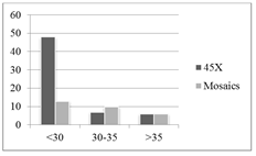
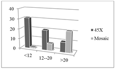
A database search between August 2010 to April 2018 identified 90 cases of TS after karyotyping. The age of pregnant women ranged from 19 to 40 yrs. and gestational age (GA) 9 to 32 weeks. The USG findings were retrieved and the most common features were noted and tabulated. Table 2 shows the range of maternal age and fetal karyotypes, done from either AC or CVS. Interestingly, the average maternal age of the TS fetuses with a mosaic was 31 years (range 25-40 years), whereas among the mothers of TS fetuses with 45, X, it was 28 years (range 19–40 years). We noted the higher number of diagnosis (61/90, 68%) in the <30 age groups as compare to (17/90, 19%) in 30-35 yr and (12/90, 13%) in >35yr age groups. More TS were diagnosed in the <12w GA period (32) compare to advanced age groups, 17 fetuses for 30-35yr and 12 for >35 yr. The 45X karyotype was found in 61 cases (67.7%; 61/90) and the other 29 cases (32.2%; 29/90) were mosaics (Table 3). Detection for 45X was higher in <12 weeks (50.8%, 31/61) period while less in later stage (33%, 16% respectively). However detection for mosaic was higher after 20weeks (72.4%, 21/29) compare to first trimester (3%, 24% respectively).
|
Age Group (years) |
Number of Fetus |
45X |
Mosaic |
|
<30 |
61 |
48 |
13 |
|
30-35 |
17 |
7 |
10 |
|
>35 |
12 |
6 |
6 |
Table 1: Number of fetus and karyotypes as function of women age groups.
|
Gestation age (week) |
Number of Fetus |
45X |
Mosaic |
|
<12 |
32 |
31 |
1 |
|
12-20 |
27 |
20 |
7 |
|
>20 |
31 |
10 |
21 |
|
Total |
90 |
61 |
29 |
Table 2: Number of fetus and karyotypes as function of gestation age in weeks.
3.2 Karyotypes and ultrasound findings
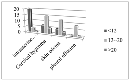
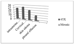
85 fetuses (94.4%; 85/90) showed symptoms on sonography (Table 4) which were more or less similar and could be grouped, whereas in 5 cases (5.6%; 5/90) other abnormalities were identified, not tabulated here. A substantial proportion of fetuses with TS showed early-onset signs that could be detected in the first trimester <12 weeks. We notice here that none of the mosaic TS fetuses (0%, 0/29) showed any abnormal structural features suspicious of TS, and were diagnosed later by karyotyping. 25 fetuses were lost as intrauterine fetal demise (IUD), of which 20 fetuses (80%, 20/25, p<0.001) in early trimester compare to later period (20%, 5/25). 24 of IUD were 45X karyotype (96%, 24/25, p<0.001) against one of mosaic karyotype (4%, 1/25).
|
Karyotype |
Intrauterine Death |
Cystic Hygroma |
Skin edema |
Cavity Effusion |
|
45X |
24 |
27 |
21 |
11 |
|
Mosaic |
1 |
0 |
0 |
0 |
|
P |
<0.001 |
<0.001 |
<0.001 |
<0.001 |
Table 3: Ultrasound findings and number of fetuses and as function of karyotypes.
|
Gestation age (weeks) |
Intrauterine Death |
Cystic Hygroma |
Skin edema |
Cavity Effusion |
|
<12 |
20 |
7 |
5 |
1 |
|
12-20 |
4 |
15 |
13 |
9 |
|
>20 |
1 |
5 |
3 |
1 |
|
Total |
25 |
27 |
21 |
11 |
|
P |
<0.001 |
<0.001 |
<0.001 |
<0.001 |
Table 4: Number of fetus with ultrasound findings as function of gestation age in weeks.
This demonstrates that 45X are spontaneously lost in early trimester. Among the congenital anomalies detected by sonography on fetuses with TS, Cystic hygroma was the most frequently seen (31.8%; 27/85) of which most were diagnosed in the second trimester (55.6%; 15/27) and only in 45X karyotype (100%, 27/27, p<0.001). Skin edema was seen in 21 fetuses (11.6% 21/90) of the 90 cases also limited to the 45X karyotype fetuses (100%, 21/21, p<0.001) as was pleural effusion in 11 fetuses (12%, 11/90) limited to 45X karyotype (100%, 11/11, p<0.001). Other 5 cases included congenital heart defects diagnosed by sonography in 2 fetuses (2.2%), 1 case of oligohydramnios (1.1%), one case with abnormally short femur length (1.1%) and one case of diaphragmatic hernia (1.1%) from the 90 cases.
3. Discussion
The detection of TS was higher in the early reproductive age women (<30yr) as most cases were detected accidentally at antenatal exams by 11 weeks since there were no routine genetic screenings for fetal chromosomal abnormalities at early GA,. Early pregnancy USG is not routinely recommended in uncomplicated and asymptomatic pregnancies. However for AMA, prenatal genetic testing was routinely performed and women were evaluated more regularly to monitor viability of intrauterine embryo. AMA become a risk factor for chromosomal abnormalities, including TS, owing to nondisjunction phenomenon that occurs in the period of meiosis during maternal oogenesis and few studies demonstrated a higher occurrence of TS of monosomy X karyotype rather than mosaic karyotypes [4, 5]. This accounts for majority cases (61, 68%) in <30 yr group but less cases (17, 18%) in 30-35 yr and (12, 13%) in >35 yrs groups of our study group. Of those detected in AMA, most are likely to be mosaic that had no, or minimal, sonographic features for early trimester detection. In the recent years, the yearly incidence has decreased owing to an improved diagnostic accuracy both by karyotyping and USG.
As for gestational age (GA), we noticed there was more or less same total number of TS fetuses detection, or 32, 27 and 31, for the <12 wks, 12-20 wks and >20 wks GA respectively. From these data, it appears that detection is same throughout pregnancy but after karyotyping, we have a different view. Comparing the detection of classic to mosaic in each groups, we noticed in the <12 weeks, the 45X made up the majority (31/32, 97%) against one mosaic (1/32, 3%). For the 12-20 weeks, of 27 TS fetuses, 20 (74%) were classic 45X while 7 (26%) mosaic. However in the >20 weeks, of 31 TS fetuses 21 (68%) were mosaic while 10 (32%) were 45X. We deduced that most of the diagnosis in the early trimester were based on sonographic findings before karyotyping confirmation. That says, the 45X monosomy produces enough cluster of findings in early trimester for diagnosis of TS. In the >20 weeks period the detection of 45X is much less because either they have already been diagnoses or simply fetus underwent spontaneous abortion.

Figure 1: Number of fetus and karyotypes as function of mother’s age groups.

Figure 2: Number of fetus and karyotypes as function of gestation age in weeks.
The detection of mosaic in later trimester accounts for the fact that they were detected while screening for other condition, AMA, or minimal features suspicious of TS. From these graphs we conclude that most severe phenotypes are in the 45X, which apparently produced enough sonographic features for diagnosis TS but this is not the case for the mosaic condition. This emphasizes the importance of karyotyping for final diagnosis of TS. Therefore USG can miss TS fetus in mosaic phenotypes since absence of feature may allow karyotypically TS fetus to go unnoticed throughout pregnancy. Prenatal diagnosis of TS can be highly variable depending on the degree of anomalies present is associated with a high rate of fetal loss, with an estimated greater than 90% of fetuses not surviving to term. Of 25 (25/90, 27%) intrauterine death, only one (1/25, 4%) was mosaic. The other 24 (96%, p<0.001) classic 45X were all lost in <12 weeks. Most of these fetuses are spontaneously aborted, medically terminated and only a few will persist throughout pregnancy accounting for low prevalence of. 45X TS postnatal. The incidence of mosaic postnatal is higher as the lack of USG features usually results in “falsely” diagnosed normal fetus. Cystic hygroma 27 (30%), skin edema 21 (23%), and pleural effusion 11 (12%) were the common sonographic markers. These ultrasonography findings; have therefore shown a statistically significant impact (p<0.001) for the prenatal ultrasound diagnosis of TS. The expertise of the sonographer is important for detecting these findings. About 96% of 45X fetuses are spontaneously lost in early trimester as per our study. Of those surviving to second trimester, almost 100% will express one or more of the cluster of findings while the mosaic fetuses hardly express any, (p<0.001). Clinical significance is that 45X fetuses are diagnosed in early trimester, <12 weeks, while the mosaic fetuses later in 2nd-3rd trimester, >20 weeks, or even postnatal, (p<0.001). Clinically the cluster of ultrasonography findings are highly predictive of TS, whenever present, (p<0.001) while absence does not rule out TS but their presence are also dependent on karyotype of fetus. Hence a marked significance in term of fetuses karyotypes, 45X against mosaic (100% vs. 0%, p<0.0001) for features appearance.

Figure 3: Ultrasound findings in fetus as function of gestation age in weeks.

Figure 4: Ultrasound findings in fetus as function of karyotypes.
When fetuses with TS grow, there is a developmental delay or hypoplasia of the lymphatic system and lymphatic drainage to venous system. In the normal condition, fetal lymphatic vessels drain in sacs lateral to jugular veins in neck. In TS fetuses, defect in the communication of lymphatic and venous system results in accumulation of fluid in the upper dermis of the nuchal skin that consequently expands to form the characteristic cystic hygroma appearance [6, 7]. Though most fetuses will end in spontaneous abortion, of those surviving beyond the first trimester, there may be spontaneous resolution of CH who at birth may present with webbed neck [8]. Fetal hydrops is also a feature of TS fetuses. Even if the accumulation of fluid in the pleural cavity, either unilateral or bilateral, has been reported in TS fetuses, we cannot say it is a marker specific to TS as other condition, including lung lesions, prenatal infections and fetal heart defects,.may also present with pleural effusion [9] Along the course of fetal development, organogenesis occurs and deformities of organ, if any, or gravity, or progression, of already established anomalies can be monitored by sonographer.
The significance of our findings combined with evidence of previous reported studies is that as early as 11 weeks on routine antenatal USG, an abnormal thickening of nuchal translucency (NT) can suggest TS in 30-70% [10, 11], and the “syndromic marker are highly suggestive of TS.. Multiple studies have been published to which our study results are in conformity to. A study by Brooke Polivka based on case reports had similar findings [12] and another extensive study consisting of 69 TS fetuses confirmed similar findings and also confirm that most of the findings were in the classic 45X rather than the mosaic TS fetuses [13]. As we approach the second trimester, more syndromic features will appear that may consolidate USG diagnosis of TS such as abnormal thickening of NT, delayed nasal bone ossification, abnormal ductus venosus velocity waveform, congenital heart defects, fetal hydrops (non-immune) and other findings of our study and these cluster of sonographic markers are highly predictive of TS in early pregnancy [14, 15]. However it is worth to note that the absence of feature cannot rule out TS. The mosaic phenotype may be completely asymptomatic. Whenever there is a suspicion of TS, karyotyping is the best option and remains by far the gold standard for diagnosing TS. In so doing, withing limit of experimental errors, we confirmed that the clusters of ultrasonography findings are of fundamental clinical significance for the prenatal ultrasound diagnosis of fetuses.
In short, the cluster of ultrasonography findings; intrauterine death, cystic hygroma, skin edema and cavity effusion showed a statistically significant impact (p<0.001) for the prenatal ultrasound diagnosis of TS. About 96% of 45X fetuses are spontaneously lost in early trimester. Of those surviving to second trimester, almost 100% will express one or more of the cluster of findings while the mosaic fetuses hardly express any, (p<0.001). The clinical significance is that 45X fetuses are diagnosed in early trimester while the mosaic fetuses later in 2nd-3rd trimester or even postnatal, (p<0.001). The mosaic fetuses can go unnoticed throughout pregnancy defying sonographer expertise by mimicking normal fetuses only to be diagnosed later in childhood, for short stature, or in adulthood, for infertility. Clinically the cluster of ultrasonography findings are highly predictive of TS, whenever present, (p<0.001) while absence does not rule out TS but their presence are also dependent on karyotype of fetus. Hence, though these markers are clinically significant, karyotyping remains the gold standard for definitive diagnosis of TS. The overall prognosis of TS is variable and highly dependent on the associated anomalies and the severity of the anomalies. The earlier the diagnosis the better it is, for parent-to-be to decide on the current pregnancy. If Turner syndrome is diagnosed after birth until adulthood, there are updated recommendations for care and management.
Conflict of Interest
The authors have no conflict of interest.
References
- Gravholt CH, Backeljauw P. New international Turner syndrome guideline: A multi-society feat. European Journal of Endocrinology 177 (2017): 1-2.
- Gravholt CH, Andersen NH, Conway GS, et al. Clinical practice guidelines for the care of girls and women with Turner syndrome. Proceedings from the 2016 Cincinnati International Turner Syndrome Meeting. Eur J Endocrinol 177 (2017): 1-70.
- Marcus A Tuke, Andrew R Wood, Robin N Beaumont, et al. Mosaic Turner syndrome shows reduced penetrance in an adult population study. Genetics in Medicine (2018).
- Hagman A, Wennerholm UB, Kallen K, et al. Women who gave birth to girls with Turner syndrome: maternal and neonatal characteristics. Human Reproduction 25 (2010): 1553-1560.
- Kim YJ, Lee JE, Kim SH, et al. Maternal age-specific rates of fetal chromosomal abnormalities in Korean pregnant women of advanced maternal age. Obstet Gynecol Sci 56 (2013): 160-166.
- von Kaisenberg CS, Nicolaides KH, Brand-Saberi B. Lymphatic vessel hypoplasia in fetuses with Turner syndrome. Human Reproduction 14 (1999): 823-826.
- von Kaisenberg CS, Wilting J, Dork T, et al. Lymphatic capillary hypoplasia in the skin of fetuses with increased nuchal translucency and Turner’s syndrome: comparison with trisomies and controls. MHR: Basic science of reproductive medicine 16 (2010): 778-789.
- Mostello DJ, Siddiqi TA. Spontaneous resolution of fetal cystic hygroma and hydrops in Turner syndrome. Obstet Gynecol 73 (1989): 862-865.
- Kosinski P, Abramowicz JS. Early 2D/3D ultrasound diagnosis of pleural effusion in fetuses with Turner syndrome. J Clin Ultrasound 46 (2018): 585-587.
- Alpman A, Akgul M, Arikan EA, et al. Prenatally diagnosed Turner syndrome and cystic hygroma: incidence and reasons for referrals. Fetal Diagn Ther 25 (2009): 58-61.
- Alexioy E, Trakakis E, Kassanos D, et al. Predictive value of increased nuchal translucency as a screening test for the detection of fetal chromosomal abnormalities. J Matern Fetal Neonatal Med 22 (2009): 857-862.
- Brooke Polivka, Kelsy L Merideth. Sonographic Prenatal Diagnosis of Turner Syndrome. Journal of Diagnostic Medical Sonography 31 (2014): 99-102.
- Csaba Papp, Artur Beke, Gabor Mezei, et al. Prenatal Diagnosis of Turner Syndrome. journal of ultrasound in medicine 25 (2006): 711-717.
- Bronshtein MZE, Blazer S. A characteristic cluster of fetal sonographic markers that are predictive of fetal Turner syndrome in early pregnancy. Am J Obstet Gynecol 188 (2003): 1016-1020.
- Wiechec MKA, Nocun A, Wiercinska E, et al. What are the most common first-trimester ultrasound findings in cases of Turner syndrome? J Matern Fetal Neonatal Med 30 (2017): 1632-1636.
