Chronic Racemic and (S)-Methadone Treatment Reduces hERG Gene Expression in Human Primary Cardiomyocytes
Article Information
Noor Fatmawati Mokhtar* and Mohd Farid Hussin
Institute for Research in Molecular Medicine (INFORMM), Universiti Sains Malaysia, Health Campus, 16150 Kubang Kerian, Kelantan, Malaysia
*Corresponding Author: Noor Fatmawati Mokhtar, Institute for Research in Molecular Medicine (INFORMM), Universiti Sains Malaysia, Health Campus, 16150 Kubang Kerian, Kelantan, Malaysia, Phone: 6097672411; Fax: 6097648673;
Received: 21 November 2017; Accepted: 27 November 2017; Published: 29 November 2017
Share at FacebookAbstract
Background: Methadone is now increasingly being used to control opioid withdrawal symptoms, and recently, chronic pain. With increasing reports of cardiac problems induced by methadone, we aimed to determine effect of methadone on hERG expression after chronic treatment with racemic and the two enantiomers (S- and R-) of methadone. The molecular mechanism underlying methadone-disrupted hERG expression in human primary cardiomyocytes was also investigated.
Methods: Cardiomyocytes were treated for 5 days with racemic-, (S)- and (R)-methadone at 3, 2, and 7 μM, respectively. The effect of racemic-, (S)-, and (R)-methadone on hERG, SGK3, and Hsp90 mRNA expression was examined by real-time PCR. Protein expression of hERG was determined by western blotting.
Results: Chronic treatment with racemic- and (S)-methadone but not (R)-methadone, significantly suppressed hERG mRNA expression and down-regulated SGK3 mRNA expression. However, hERG protein expression was only affected by chronic (S)-methadone treatment. Hsp90 mRNA expression was not affected by any form of methadone. Conclusions: We conclude that after chronic treatment, racemic- and (S)-methadone have similar potencies in reducing hERG mRNA expression, and this act via disruption of SGK3 expression.
Keywords
Racemic Methadone; (S)-Methadone; (R)-Methadone; Chronic Treatment; Herg Expression; Cardiomyocytes
Article Details
1. Background
Methadone was first introduced by the United States Public Health Service Hospital in the 1960s as a treatment for opioid abstinence syndrome, and still remains the most widely prescribed and most efficacious treatment for this condition [1]. More recently, methadone is used in palliative care to control chronic cancer pain [2]. Due to its low production cost, fast-acting and long-lasting effect, methadone usage is steadily increasing [3]. The drug is currently administered as a racemic form, composed of a mixture of (R)- and (S)-enantiomers, although (R)-methadone is reported to be primarily responsible for its therapeutic benefit, as racemic-methadone produces a similar therapeutic outcome [4]. (S)-methadone is primarily responsible for off-target, adverse effects [5].
The human ether-a-go-go-related gene (hERG) encodes a potassium channel that has a critical role in normal heart function. Recent advances have shown that compared with other K+ channels, hERG-encoded channels are more susceptible, and extraordinarily sensitive to, drug blockade [6]. Blocking hERG channels causes the drug-induced form of long QT syndrome (LQTS), a cardiac disorder that carries an increased risk of cardiac arrhythmias and sudden death. Therefore, hERG has generated considerable interest in pharmacologically linked arrhythmias.
There has been a significant increase in cases of QT interval prolongation and torsade des pointes (TdP) associated with a wide range of methadone dosages, including those recommended for methadone maintenance treatment and palliative care [7-9]. With growing concern that chronic methadone treatment can lead to future health problems and major complications, the effect of methadone on hERG needs to be determined.
Many in vitro studies have found that methadone inhibits hERG channels with IC50 values ranging between 1.7 and 20 µM [10-15]. Further in vitro studies have shown methadone-induced QT interval prolongation in isolated perfused rabbit heart [16] and action potentials from human stem-cell derived cardiomyocytes [14].
Many previous in vitro studies examining drug effects on hERG have used the patch-clamp method to determine acute effects in a hERG-expressing heterologous system e.g., HEK293-hERG [12, 15]. However, for some drugs, hERG blockade may not be detected at the acute treatment stage, but may still affect hERG expression at clinically relevant concentrations. Example of these include arsenic trioxide (anti-cancer), pentamidine (anti-infective/microbial), probucol (cholesterol-lowering drug), and digitoxin (cardiac glycoside), which have all been retracted from the market due to their effect on reducing hERG expression [17-20]. Others such as ketoconazole (anti-fungal) and fluoxetine (anti-depressant) acutely block hERG currents and simultaneously disrupt its expression [21, 22]. Notably, growing evidence highlights that defective hERG expression also leads to QT interval prolongation with increased predisposition to cardiac arrhythmias, TdP, and sudden cardiac arrest [23, 24].
All of these observations on drug-induced alterations in hERG expression are similar to biogenesis defects implicated in the vast majority of examples of hERG mutation-mediated QT prolongation [25]. Serum and glucocorticoid inducible kinase-3 (SGK3) and the 90-kDa heat shock protein (Hsp90) regulate cardiac hERG expression and may be involved in hERG biogenesis defects [26, 27]. Previously, SGK3 and hERG coexpression in Xenopus oocytes was shown to enhance steady-state currents and cell membrane protein abundance [27]. Hsp90 is an abundant molecular chaperone intimately involved in protein folding, activation, and stabilization [28]. Hsp90 inhibitors e.g., geldanamycin, disrupt the Hsp90-ATPase cycle and suppress hERG protein maturation, facilitating its degradation via the ubiquitin-proteasome system [29].
In this study, we investigated the effect of chronic treatment of racemic- and the two (S- and R-) methadone enantiomers on hERG gene and protein expression in a native cardiomyocyte model system. We also examined SGK3 and Hsp90 mechanisms in methadone disruption of hERG expression.
2. Methods
2.1 Cell culture
Human cardiac myocytes (HCMs), cardiac myocyte medium, cardiac myocyte growth serum, fetal bovine serum, and penicillin-streptomycin antibiotic were purchased from ScienCell Research Laboratories (Carlsbad, California, USA). Cells were grown on 10-mm or 35-mm Falcon culture dishes (Becton Dickinson, Franklin Lakes, New Jersey, USA) coated with poly-L-lysine (Sigma-Aldrich,St. Louis, Missouri, USA), and allowed to reach approximately 90% confluency before passaging for experiments. Experiments were performed in 35-mm culture dishes with initial cell densities of 2 ´ 105 and 4 ´ 105 cells/dish for RNA and total protein extraction, respectively. TrypLETM Select was used for passaging HCMs, according to the manufacturer’s instruction (Life Technologies, Grand Island, New York, USA). All cultured cells were maintained in a humidified incubator at 37°C, 100% relative humidity, and with 5% regulated CO2.
2.2 Pharmacology
Three types of methadone were used throughout the study: racemic-, (R)- and (S)- methadone (Toronto Research Chemicals, Toronto, Ontario, Canada). For each type, a single concentration was used, based on published patch-clamp data [12], and corresponding to the IC50 (i.e., concentration that blocks 50% of hERG currents): 3, 2, and 7 µM, respectively. Stock concentrations (1 mM) were prepared in distilled water and stored at ?20°C until needed.
2.3 Quantitative and semi-quantitative real-time PCR
Total RNA was extracted from cell dishes at approximately 80% confluency using Trizol, according to the manufacturer’s instructions (Invitrogen, Grand Island, New York, USA). cDNA was synthesized from 1 µg total RNA using the QuantiTect Reverse Transcription Kit (Qiagen, Venlo, Netherlands). The resulting cDNA was stored at -20°C until use. Real-time PCR was performed using SYBR Green PCR mix (Qiagen) with an ABI 7000 Prism real-time PCR machine (Applied Biosystems, Grand Island, New York, USA).
The cycling conditions were: initial PCR activation at 95°C for 5 min, then 40 cycles of denaturation at 60°C for 10 sec and combined annealing/extension at 60°C for 30 sec, followed by a thermal dissociation protocol (Bioline, London, UK). The primers (1st BASE, Selangor, Malaysia) used were: hERG, 5??TGCTCATTGGCTCCCTCATGTATG?3¢ (F) and 5¢?GCATCTGTGTGTGGTAGCGG?3¢ (R), amplicon size 99 bp; troponin I, 5¢?CCAACTACCGCGCTTATGC?3¢ (F) and 5¢?CTCGCTCCAGCTCTTGCTTT?3¢ (R), amplicon size 120 bp; ?-actin, 5¢-ATTGCCGACAGGATGCAGAAG? 3¢ (F) and 5¢? TAGAAGCATTTGCGGTGGACG?3¢ (R), amplicon size 203 bp; SGK3, 5¢?TGTGGGACACCAGAGTATCTTG?3¢ (F) and 5¢?AAGGAGGCAATCCATACAGCA?3¢ (R), amplicon size 118 bp; Hsp90, 5¢?GACCAGAAATCCCGACGAT?3¢ (F) and 5¢?AAATGCTTCACTGCCAAGTG?3¢ (R), amplicon size 96 bp. hERG mRNA expression in treated and untreated control samples was normalized to expression of the housekeeping gene, ?-actin. Relative hERG expression using ?-actin as the reference gene was calculated based on the threshold cycle number (CT) of each PCR, according to the Pfaffl method [30]. hERG mRNA expression levels were expressed as fold-difference of levels in treated versus control samples normalized by ?-actin.
2.4 Western blotting
Total protein was extracted in RIPA buffer, according to the manufacturer’s protocol (Thermo Scientific, Grand Island, New York, USA). Total protein concentration was determined using a standard curve prepared with known concentrations of bovine serum albumin (Sigma-Aldrich). Samples were then prepared for loading onto gels or stored at ?20°C. For hERG protein detection, total protein extracts were separated on SDS polyacrylamide gels and electrophoretically transferred onto nitrocellulose membranes (Biorad, USA). Bands were detected by chemiluminescence (Nacalai Tesque, Kyoto, Japan).
A primary hERG antibody (Sigma-Aldrich) and anti-rabbit HRP-conjugated secondary antibody (GenScript, Piscataway, New Jersey, USA) were used accordingly. To determine equal protein loading, ?-actin (Santa Cruz Biotechnology Inc., Dallas, Texas, USA) was used. To improve the color, sharpness, and contrast of western blot images, the Format Picture setting in Microsoft Word 2010 was used. Graphical representation of hERG protein expression was quantified using Image J (http://imagej.nih.gov/ij/NIH) and normalized to the ?-actin loading control
2.5 Data analysis
Data are presented as mean ± standard error of the mean (SEM) with n = 3. Statistical significance was determined using Student’s t-tests.
3. Results
3.1 Reduced hERG mRNA expression after chronic treatment with racemic- and (S)-methadone
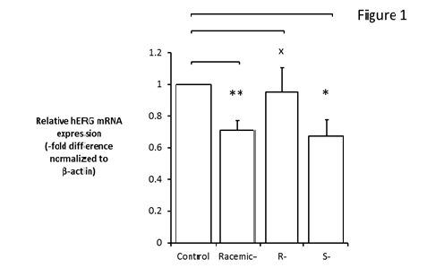
After 5 days of treatment with IC50 racemic-, (R)- and (S)- methadone, relative real-time PCR was performed to measure hERG gene expression. The CT values for each sample were normalized to the CT value of ?-actin, as a reference gene. Relative hERG mRNA expression was significantly reduced by racemic- and (S)-methadone after 5 days of treatment (P < 0.05), but was not significantly affected by (R)-methadone (Figure 1).
Cardiomyocytes were treated with racemic-, (S)- and (R)-methadone for 5 days, and then total RNA extracted and cDNA synthesized. Relative hERG gene expression was determined by real-time PCR based on the Pfaffl method [30]. Data was collected from n = 3 independent experiments (*P < 0.05). X indicates no significant differences.
3.2 Reduced hERG total protein expression after chronic treatment with racemic and (S)-methadone
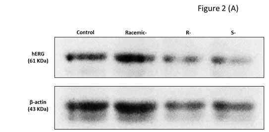
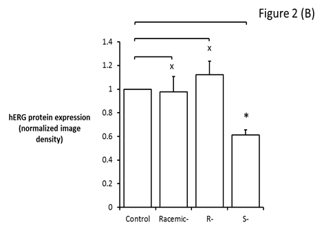
Western blotting was performed to investigate the effect of IC50 racemic-, (R)- and (S)- methadone on hERG protein expression in human primary cardiomyocytes. We observed significantly decreased hERG protein expression after 5 days of treatment with (S)-methadone (P < 0.05), but not racemic- or (R)-methadone (Figure 2).
Figure 2: Reduced hERG protein expression in primary human cardiomyocytes after 5 days of treatment with (S)-methadone.
Cardiomyocytes were treated with racemic-, (S)- and (R)-methadone for 5 days, and then total protein extracted. (A) Western blotting using a hERG primary antibody (1:1000) and HRP-conjugated secondary antibody (1:10000). ?-actin primary antibody (1:1000) was also used.
3.3 Modulation of the transcription factor SGK3, but not Hsp90, by racemic- and (S)-methadone
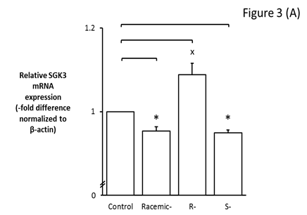
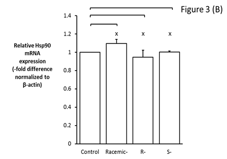
Because racemic- and (S)-methadone significantly affect hERG gene and/or protein expression, the molecular mechanism for this disruption was further investigated by examining expression of the transcription factor, SGK3, and a chaperone protein, Hsp90. SGK3 gene expression was significantly reduced by IC50 racemic- and S-methadone (P < 0.05) (Figure 3A), with no significant effect of R-methadone observed. However, Hsp90 gene expression was not affected by chronic treatment with racemic-, (R)-, and (S)-methadone (Figure 3B).
Figure 3: Modulation by racemic-, (R)- and (S)-methadone of SGK3 but not Hsp90.
Cardiomyocyte cDNA after 5 days treatment with racemic- and (S)-methadone was examined by real-time PCR for SGK3 and Hsp90 expression. (A) Graphical representation of SGK3 mRNA expression normalized to ?-actin. SGK3 gene expression was significantly reduced after chronic treatment with IC50 racemic- and (S)-methadone.
Figure 3: (B) Graphical representation of Hsp90 mRNA expression normalized to ?-actin. Hsp90 gene expression was not affected by chronic treatment with racemic-, (R)-, and (S)-methadone. Relative gene expression was measured according to the Pfaffl method [30]. Data are representative of n = 3 independent repeats with X indicates no significant differences (P > 0.05).
4. Discussion
Although methadone has been established as a safe drug, reports of methadone-associated arrhythmia have increased substantially from 1997-2011, with the number of cases of QT interval prolongation and TdP similar to that of other highly proarrhythmic drugs, such as dofetilide [31]. Consequently, there is an urgent need to reassess the effect of methadone on cardiac health.
New evidence is emerging that altered plasma membrane expression of hERG leads to QT interval prolongation with increased predisposition to cardiac arrhythmia and sudden cardiac arrest [23, 24]. Chronic treatment with certain clinically approved drugs, e.g., arsenic trioxide (anti-cancer), pentamidine (anti-infective/microbial), probucol (cholesterol-lowering drug), digitoxin (cardiac glycoside), ketoconazole (anti-fungal), and fluoxetine (anti-depressant), disrupts hERG expression and can potentially lead to LQTS [17-22]. In the worst cases, these findings have resulted in drug withdrawal from the market.
Here, at a concentration that has been reported to block 50% of hERG channels (IC50), hERG mRNA expression in human cardiomyocytes was significantly reduced after 5 days treatment with racemic- and (S)-methadone, but not (R)-methadone (Figure 1). Thus, it appears that the disruptive effect of (S)-methadone on hERG gene expression is dominantly in the racemic-form.
Altered hERG gene expression by IC50 (S)-methadone is corroborated by our protein analysis, with hERG protein expression also significantly reduced by (S)-methadone and not (R)-methadone (Figure 2). These results are in accordance with two independent studies showing that (S)-methadone is more potent against hERG-encoded channels than (R)-methadone [11, 12]. However, we did not observe the disruptive effect of (S)-methadone on hERG protein expression in the racemic-form (Figure 2), supporting the observations that racemic-methadone produces a similar therapeutic outcome compared with (R)-methadone alone [4]. Nevertheless, replacing racemic- with (R)-methadone in patients receiving maintenance treatment has been shown to reduce QT interval prolongation [5].
Essentially, the variation/ratios of (R)- and (S)-methadone in the racemic-form may be influencing the therapeutic response [33]. However, there is still inadequate data to show this unequivocally. Furthermore, the magnitude of the effect also depends on an individual’s predisposition in drug clearance and interactions [34].
To further understand the molecular mechanisms of racemic- and (S)-methadone, we examined the involvement of SGK3 and Hsp90. These molecules are implicated in hERG gene transcriptional regulation and protein trafficking to the cardiac plasma membrane, respectively [26, 17]. Altered expression of both molecules in the mechanism of drug-induced hERG expression defects has been reported elsewhere [24]. We found significantly reduced SGK3 gene expression after 5 days treatment with racemic- and (S)-methadone, corroborating studies demonstrating transcriptional regulation of SGK3 on hERG [26, 27].
hERG abundance in the plasma membrane is a key determinant of hERG functionality. Maturation and trafficking of hERG protein to the plasma membrane is dependent upon chaperone proteins, such as Hsp90 [17, 26]. With the exception of arsenic oxide, the mechanism responsible for reduced trafficking remains unknown, therefore we wanted to examine a potential trafficking mechanism for methadone. We found no significant changes in Hsp90 gene expression with racemic-methadone or either of its isomers. This implies that reduced hERG protein expression following chronic methadone treatment is not regulated by Hsp90. However, in order to ascertain that Hsp90 does not play a role in methadone-reduced hERG expression, further studies must be performed to examine Hsp90 protein levels.
5. Conclusions
Our findings conclude that chronic racemic- and (S)-methadone, but not (R)-methadone treatment reduces hERG gene expression in human cardiomyocytes, and this involves interruption of transcription factor SGK3 gene expression. Although hERG protein was also affected by chronic (S)-methadone treatment, Hsp90 which plays role in protein maturation and trafficking is not affected by any methadone forms. More work is needed to understand these methadone-induced alterations in hERG expression, and develop further strategies for determining cardiac risk for this drug in which a consideration to prescribe only the (R)-methadone for any treatment is highly recommended.
6. List of abbreviations
hERG: Human Ether-a-go-go-Related Gene
LQTS: Long QT Syndrome
TdP: Torsade Des Pointes
HCMs: Human Cardiac Myocytes
Hsp90: Heat Shock Protein
SGK3: Serum and Glucocorticoid Inducible Kinase-3
7. Declarations
Ethics approval and consent to participate
Not applicable
Consent for publication
Not applicable
Availability of data and materials
Data and materials related to this work are available upon request.
Competing interests
The authors declare that they have no competing interests
Funding
This work was supported by the Ministry of Education, Malaysia and Universiti Sains Malaysia under the Incentive and Short Term Grant (No. 304/CIPPM/61312013).
Author’s contribution
NFM is the study’s principal investigator and was involved in the initial and final drafting of the manuscript’s intellectual content. MFH was responsible for the experimental work, data collection, and statistical analysis. Both authors have read and approved the manuscript.
Acknowledgements
We thank Ministry of Education, Malaysia and Universiti Sains Malaysia for funding supports. We also thank Institute for Research in Molecular Medicine (INFORMM) for the availability of research equipment and facilities.
References
- Center for Substance Abuse Treatment. Medication-Assisted Treatment for Opioid Addiction in Opioid Treatment Programs. Treatment Improvement Protocol (TIP) Series 43. HHS Publication No. (SMA) 12-4214. Rockville, MD: Substance Abuse and Mental Health Services Administration. 2005.
- Leppert W. The role of methadone in cancer pain treatment--a review. Int J Clin Pract 63 (2009): 1095-109.
- Substance Abuse and Mental Health Services Administration, Center for Behavioral Health Statistics and Quality. The N-SSATS Report: Trends in the Use of Methadone and Buprenorphine at Substance Abuse Treatment Facilities: 2003 to 2011. Rockville, MD. 2013.
- Mccance-Katz EF. (R)-methadone versus racemic methadone: what is best for patient care? Addiction 106 (2011): 687-688.
- Ansermot N, Albayrak O, Schläpfer J, Crettol S, Croquette-Krokar M, Bourquin M, et al. Substitition of (R,S)-methadone by (R)-methadone: impact on QTc interval. Arch Intern Med 170 (2008): 529-536.
- Mitcheson JS. hERG potassium channels and the structural basis of drug-induced arrhythmias. Chem Res Toxicol 21 (2008): 1005-1010.
- Stringer J, Welsh C, Tommasello A. Methadone-associated Q-T interval prolongation and torsades de pointes. Am J Health Syst Pharm 66 (2009): 825-833.
- Wilcock A, Beattie JM. Prolonged QT interval and methadone: implications for palliative care. Curr Opin Support Palliat Care 3 (2009): 252-257.
- Hanon S, Seewald RM, Yang F, Schweitzer P, Rosman, J. Ventricular arrhythmias in patients treated with methadone for opioid dependence. J Interv Card Electrophysiol 28 (2010): 19-22.
- Katchman AN, Mcgroary KA, Kilborn MJ, Kornick CA, Manfredi PL, Woosley RL et al. Influence of opioid agonists on cardiac human ether-a-go-go-related gene K+ currents. J Pharmacol Exp Ther 303 (2002): 688-694.
- Kornick CA, Kilborn MJ, Santiago-Palma J, Schulman G, Thaler HT, Keefe DL et al. QTc interval prolongation associated with intravenous methadone. Pain 105 (2003): 499-506.
- Eap CB, Crettol S, Rougier JS, Schläpfer J, SintraGrilo L, Déglon JJ, et al. Stereoselective block of hERG channel by (S)-methadone and QT interval prolongation in CYP2B6 slow metabolizers. Clin Pharmacol Ther 81 (2007): 719-728.
- Lin C, Somberg T, Molnar J, Somberg J. The effects of chiral isolates of methadone on the cardiac potassium channel Ikr. Cardiology113 (2009): 59-65.
- Kuryshev YA, Bruening-Wright A, Brown AM, Kirsch GE. Increased cardiac risk in concomitant methadone and diazepam treatment: pharmacodynamic interactions in cardiac ion channels. J Cardiovasc Pharmacol 56 (2010): 420-430.
- Zünkler BJ, Wos-Maganga M. Comparison of the effects of methadone and heroin on human ether-à-go-go-related gene channels. Cardiovasc Toxicol 10 (2010): 161-165.
- Katchman AN, Koerner J, Tosaka T, Woosley RL, Ebert SN. Comparative evaluation of HERG currents and QT intervals following challenge with suspected torsadogenic and nontorsadogenic drugs. J Pharmacol Exp Ther 316 (2006): 1098-1106.
- Ficker E, Dennis AT, Wang L, Brown AM. Role of the cytosolic chaperones Hsp70 and Hsp90 in maturation of the cardiac potassium channel HERG. Circ Res 92 (2003): e87-e100.
- Cordes JS, Sun Z, Lloyd DB, Bradley JA, Opsahl AC, Tengowski MW, et al. Pentamidine reduces hERG expression to prolong the QT interval. Br J Pharmacol 145 (2005): 15-23.
- Guo J, Massaeli H, Li W, Luo T, Shaw J, Kirshenbaum LA, et al. Identification of IKr and its trafficking disruption induced by probucol in cultured neonatal rat cardiomyocytes. J Pharmacol Exp Ther 321 (2007): 911-920.
- Wang L, Barbara AW, Wan X, Ficker E. Cardiac glycosides as novel inhibitors of human ether-a-go-go-related gene channel trafficking. J Pharmacol Exp Ther 320 (2007): 525-534.
- Takemasa H, Nagatomo T, Abe H, Kawakami K, Igarashi T, Tsurugi T, et al. Coexistence of hERG current block and disruption of protein trafficking in ketoconazole-induced long QT syndrome. Br J Pharmacol 153 (2008): 439-447.
- Hancox JC, Mitcheson JS. Combined hERG channel inhibition and disruption of trafficking in drug-induced long QT syndrome by fluoxetine: a case-study in cardiac safety pharmacology. Br J Pharmacol 149 (2006): 457-459.
- Apaja PM, Foo B, Okiyoneda T, Valinsky WC, Barriere H, Atanasiu R, et al. Ubiquitination-dependent quality control of hERG K+ channel with acquired and inherited conformational defect at the plasma membrane. Mol Biol Cell 24 (2013): 3787-3804.
- Nogawa H, Kawai T. hERG trafficking inhibition in drug-induced lethal cardiac arrhythmia. Eur J Pharmacol 741 (2014): 336-339.
- Li X, Xu J, Li M. The human ?1261 mutation of the HERG potassium channel results in a truncated protein that contains a subunit interaction domain and decreases the channel expression. J Biol Chem 272 (1996): 705-708.
- Lamothe SM, Zhang S. The serum- and glucocorticoid-inducible kinases SGK1 and SGK3 regulate hERG channel expression via ubiquitin ligase Nedd4-2 and GTPase Rab11. J Biol Chem 288 (2013): 15075-15084.
- Maier G, Palmada M, Rajamanickam J, Shumilina E, Böhmer C, Lang F. Upregulation of HERG channels by the serum and glucocorticoid inducible kinase isoform SGK3. Cell Physiol Biochem 18 (2006): 177-186.
- Peterson LB, Eskew JD, Vielhauer, GA, Blagg BS. The hERG channel is dependent upon the Hsp90? isoform for maturation and trafficking. Mol Pharm 9 (2012): 1841-1846.
- Iwai C, Li P, Kurata Y, Hoshikawa Y, Morikawa K, Maharani N, et al. Hsp90 prevents interaction between CHIP and HERG proteins to facilitate maturation of wild-type and mutant HERG proteins. Cardiovasc Res 100 (2013): 520-528.
- Pfaffl MW. Relative quantification. In: Dorak MT, editor. Real-time PCR. New York: Taylor and Francis Group; 2006 p. 63-82.
- Kao D, Bartelson BB, Khatri V, Dart R, Mehler PS, Katz D et al. Trends in Reporting Methadone-Associated Cardiac Arrhythmia, 1997?2011: An Analysis of Registry Data. Ann Intern Med 158 (2013): 735-740.
- Dennis A, Wang L, Wan X, Ficker E. hERG channel trafficking: novel targets in drug-induced long QT syndrome. Biochem Soc Trans 35 (2007): 1060-1063.
- Mitchell TB, Dyer KR, Newcombe D, Salter A, Somogyi AA, Bochner F et al. Subjective and physiological responses among racemic-methadone maintenance patients in relation to relative (S)- vs. (R)-methadone exposure. Br J Clin Pharmacol 58 (2004): 609-617.
- Crews KR, Hicks JK, Ching-Hon P, Relling MV, Evans WE. Pharmacogenomics and individualized medicine: Translating science into practice. Clin Pharmacol Ther 92 (2012): 467-475.
- Ficker E, Kuryshev YA, Dennis AT, Obejero-Paz C, Wang L, Hawryluk P, et al. Mechanisms of arsenic-induced prolongation of cardiac repolarization. Mol Pharmacol 66 (2004): 33-44.

