Case of Spontaneously Reported Hypersensitivy in the Ultra-Early Phase of Normal Tension Glaucoma in a Japanese Man
Article Information
Hisataka Fujimoto*; Junichi Kiryu
Department of Ophthalmology, Kawasaki Medical School, 577 Matsushima, Kurashiki, 701-0192 Okayama, Japan
*Corresponding author: Hisataka Fujimoto, Department of Ophthalmology, Kawasaki Medical School, 577 Matsushima, Kurashiki, 701-0192 Okayama, Japan.
Received: 04 June 2022; Accepted: 12 June 2022; Published: 16 June 2022
Citation: Hisataka Fujimoto, Junichi Kiryu. Case of Spontaneously Reported Hypersensitivy in the Ultra-Early Phase of Normal Tension Glaucoma in a Japanese Man. Journal of Ophthalmology and Research 5 (2022): 86-91.
Share at FacebookAbstract
Background: We report a case of spontaneous reported hypersensitivity in ultra-early phase in normal tension glaucoma in a Japanese professor in a medical school.
Case Presentation: At 66 years of age, the patient spontaneously noticed subjective symptoms, such as scotoma, in the visual field in the temporal lower position in the right eye while he examined his electronic microscopy (EM) study images. A slit lamp examination during the first visit to our hospital revealed a typical localized glaucomatous retinal nerve fiber layer defect in red-free filter examination and glaucomatous optic nerve head damage with 0.8 of cup-to-disc ratio and rim thinning and excavation. Moreover, optical coherence tomography examination of his optic nerve and macula (ILM-IPL/INL) revealed thinning of the retinal ganglion cell layer, especially in the right eye in which he had noticed the symptom. Furthermore, Humphrey automated perimetry was performed on the same day, confirming scotoma in the right eye coinciding with his spontaneous subjective symptom.
Conclusion and Importance: Glaucoma is normally asymptomatic and medical treatment is often delayed owing to the lack of subjective symptoms in the early phase. However, in this case, the patient’s careful and sensitive personality enabled spontaneous subjective identification in the ultra-early phase of normal tension glaucoma.
Keywords
spontaneous report, early phase, Japanese, normal tension glaucoma, glaucoma, electronic microscopy, Humphrey automated perimetry
spontaneous report articles, early phase articles, Japanese articles, normal tension glaucoma articles, glaucoma articles, electronic microscopy articles, Humphrey automated perimetry articles
Article Details
1. Intruduction
Glaucoma is the leading cause of irreversible blindness, affecting approximately 4 million people in Japan [1]. Although open angle glaucoma, including normal tension glaucoma (NTG), is considered the most common type of glaucoma in the Japanese population, 92.3% of these eyes can demonstrate intraocular pressures (IOPs) within the normal range [2]. Recently, the probability of patients with NTG progressing to unilateral blindness, and bilateral real blindness was reportedly 9.9% and 1.4% at 20 years, respectively [3]. Several large-scaled mesoscale clinical trials have investigated the natural history of untreated eyes with NTG [4,5]. A previous study demonstrated the rate of progression of visual field defects (VFDs) to vary between −0.2 and −2 dB/year [4], while another study demonstrated an average of −0.36 dB/year [5]. De Moraes et al reported that it averaged −0.33 dB/ year in their group of 81-treated NTG patients [6]. Importantly, several studies have suggested that the rate of VFD progression varies according to the patients’ eyes, distribution of glaucoma subtypes [7], and severity of VFD at presentation [8].
Herein, we report a case of ultra-early phase NTG detection in a Japanese man based on the self-examination of his electronic microscopy (EM) study images. He was a prominent professor in a medical school. His careful and perceptive personality could have promoted subjective spontaneous notification in the early phase NTG, which enabled early intervention and prevention of the visual field (VF) loss.
2. Case Presentation
The patient was a 66-year-old man with no relevant history of any systemic disease excluding a history of hypertension since 10 years. The patient was a prominent professor in a medical school. He spontaneously noticed subjective symptoms, such as scotoma in the temporal lower position in the right eye, in the VF while he examined his EM study image. The visual acuity in the right and left eye was 1.2 × -0.75 D = cyl -0.75 D Ax 70°, and was 1.2 × -2.00 D = cyl -0.75 D Ax 110°on the first refer, respectively. The IOP was also measured using a non-contact tonometer (NT-4000; NIDEK Co., Ltd, Japan), and the IOP in the right and left eye was 15 mmHg and 17 mmHg, respectively.
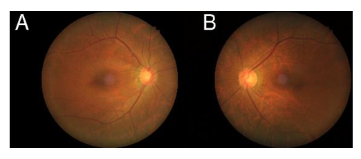
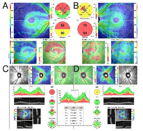
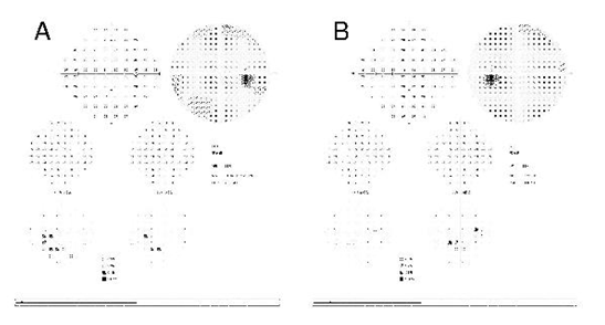
Upon slit lamp examination during the first visit of our hospital, a typical localized glaucomatous retinal nerve fiber layer defect in red-free filter examination and glaucomatous optic nerve head damage with 0.8 of cup-to-disc ratio and rim thinning and excavation were observed (Figure 1). On the same day of the examination, the bilateral optical coherence tomography (OCT) was performed. Upon the fundus OCT examination, his optic nerve and macula (ILM-IPL/INL) exhibited thinning of the retinal ganglion cell layer especially in the right eye in which he noticed the symptom (Figure 2). Moreover, Humphrey automated perimetry was performed on the same day. Early-stage scotoma in the VF in the right eye was confirmed coinciding with his spontaneous subjective symptom (Figure 3). A diagnosis of bilateral NTG was formulated. The eyes were bilaterally administered prostaglandin analogs of 0.005% Latanoprost, and successfully resulted in the IOP decrease into 14 mmHg in the right eye and 13 mmHg in the IOP in the left eye.
Figure:1 Graph illustrating the first visit to our hospital, a typical localized glaucomatous retinal nerve fiber layer defect in red-free filter examinations and glaucomatous optic nerve head damage with 0.8 of cup-to-disc ratio and rim thinning and excavation were observed both in the right eye (A) and the left eye (B)
Figure: 3 Graph illustrating the Humphrey automated perimetry performed on the same day as the examinations in Figures 1 and 2. In the Humphrey automated perimetry, early-stage scotomas were observed, especially in the right eye (A) coinciding with the patient’s spontaneous subjective symptom, and also in the asymptomatic left eye (B).
3. Discussion
Several studies have elucidated the rate of VFD progression in glaucoma patients. However, in the case of NTG, comparison of studies is challenging owing to the variations in the data according to the race, follow-up periods [12], frequency of VF testing, grade of VF, and study design concerning the inclusion criteria and research methodology. In medically treated NTG eyes, the mean rate of VFD progression reportedly varied between −0.38 and 0.02dB/year [13–16]; however, previous studies enrolled eyes with mild NTG and variable severities of VF, with the exception of advanced glaucoma.?
In initially NTG eyes, a previous study [9] reported the rate of VF progression to be −0.23 dB/year and +0.11 dB dB/year in a progressive and non-progressive group, respectively. Their findings are largely consistent with our present findings; however, our cases included both treated and non-treated NTG eyes. Thus, the subjective spontaneous notification in the early phase of NTG in the present case report enabled early intervention and the prevention of VF loss. Although the incidence of disc hemorrhage (DH), which was not observed in our patient, was previously reported to be 11–42% in eyes with NTG [17], a higher incidence of DH in our eligible population could be partly explained by the fact that the DHs are usually not observed in far-advanced glaucoma in which little of the neuroretinal rim is preserved [19]. Moreover, studies with longer follow-up periods [10] and fewer cases needing intensive therapy in our population could strengthen the reliability [20, 21].
Whether greater IOP fluctuation is a risk factor TG progression is considered controversial [22–24]. A more progressive intervention, including surgical intervention, such as for patients with advanced glaucoma, yields an unavoidable bias, especially in retrospective studies. Thus, some investigators use IOP values only prior to the development of an event when investigating the risk factors for glaucoma progression [22–24]. In summary, we demonstrated that the rate of VFD progression in the glaucoma is generally asymptomatic, thereby resulting in delayed medical treatment. However, in this case, the careful and perceptive personality of the patient enabled the subjective spontaneous notification in the ultra-early phase in NTG.
Acknowledgments:
We thank Y. Ieki and Y. Setoguchi, whose discussions, advice, and criticisms were of great benefit to this report. This study was supported in part by Research Project Grant 03B-002 from the Kawasaki Medical School (to H.F.). The author has no proprietary or commercial interest in any of the materials discussed in this article.
Conflicts of Interest:
The authors have no conflicts of interest to declare.
References
- Quigley HA, Broman AT. The number of people with glaucoma worldwide in 2010 and 2020. British Journal of Ophthalmology 90 (2006): 262-267.
- Iwase A, Araie M, Tomidokoro A, Yamamoto T, Shimizu H, et al. Prevalence and causes of low visionand blindness in a Japanese adult population: the tajimi study. Ophthalmology 113 (2006): 1354-1362.
- Sawada A, Rivera JA, Takagi D, Nishida T, Yamamoto T. Progression to legal blindness in patients with normal tension glaucoma: hospital-based study. Investigative Ophthalmology & Visual Science 56 (2015): 3635-3641.
- Anderson DR, Drance SM, Schulzer M; Collaborative Normal-Tension Glaucoma Study Group. Natural history of normal-tension glaucoma. Ophthalmology 108 (2001): 247-253.
- Heijl A, Bengtsson B, Hyman L, Leske MC; Early Manifest Glaucoma Trial Group. Natural history of open-angle glaucoma. Ophthalmology 116 (2009): 2271-2276.
- De Moraes CG, Liebmann JM, Liebmann CA, Susanna R Jr, Tello C, et al. Visual field progression outcomes in glaucoma subtypes. Acta Ophthalmol 91 (2013): 288-293.
- Heijl A, Buchholz P, Norrgren G, Bengtsson B. Rates of visual field progression in clinical glaucoma care. Acta Ophthalmologica 91 (2013): 406-412.
- Rao HL, Kumar AU, Babu JG, Senthil S, Garudadri CS. Relationship between severity of visual field loss at presentation and rate of visual field progression in glaucoma. Ophthalmology 118 (2011): 249-253.
- Miglior S, Torri V, Zeyen T, Pfeiffer N, Vaz JC, et al. Intercurrent factors associated with the development of open-angle glaucoma in the European glaucoma prevention study. American Journal of Ophthalmology 144 (2007): 266-275.
- Nakano N, Hangai M, Nakanishi H, Mori S, Nukada M, et al. Macular ganglion cell layer imaging in preperimetric glaucoma with speckle noise-reduced spectral domain optical coherence tomography. Ophthalmology 118 (2011): 2414–2426.
- Ophir A. First-visit diagnosis of preperimetric glaucoma. Open Ophthalmology Journal 4 (2010): 22-27.
- Henson DB, Shambhu S. Relative risk of progressive glaucomatous visual field loss in patients enrolled and not enrolled in a prospective longitudinal study. Archives of Ophthalmology 124 (2006): 1405-1408.
- Sakata R, Aihara M, Murata H, Mayama C, Tomidokoro A, et al. Contributing factors for progression of visual field loss in normal-tension glaucoma patients with medical treatment. Journal of Glaucoma 22 (2013): 250-254.
- De Moraes CG, Liebmann JM, Greenfield DS, Gardiner SK, Ritch R, et al. Risk factors for visual field progression in the low-pressure glaucoma treatment study. American Journal of Ophthalmology 154 (2012): 702-711.
- Ahrlich KG, De Moraes CG, Teng CC, Prata TS, Tello C, et al. Visual field progression differences between normal-tension and exfoliative high-tension glaucoma. Investigative Ophthalmology & Visual Sciences 51 (2010): 1458-1463.
- Komori S, Ishida K, Yamamoto T. Results of long-term monitoring of normal-tension Glaucoma patients receiving medical therapy: results of an 18-year follow-up. Graefes Archives for Clinical and Experimental Ophthalmology 252 (2014): 1963-1970.
- Ishida K, Yamamoto T, Sugiyama K, Kitazawa Y. Disk hemorrhage is a significantly Negative prognostic factor in normal-tension glaucoma. American Journal of Ophthalmology 129 (2000): 707-714.
- Budenz DL, Anderson DR, Feuer WJ, Beiser JA, Schiffman J, et al. Detection and prognostic significance of optic disc hemorrhages during the ocular hypertension treatment study. Ophthalmology 113 (2006): 2137-2143.
- Jonas JB, Xu L. Optic disk hemorrhages in glaucoma. American Journal of Ophthalmology 118 (1994): 1-8.
- Hendrickx KH, van den Enden A, Rasker MT, Hoyng PF. Cumulative incidence of patients with disc hemorrhages in glaucoma and the effect of therapy. Ophthalmology 101 (1994): 1165-1172.
- Miyake T, Sawada A, Yamamoto T, Miyake K, Sugiyama K, et al. Incidence of disc hemorrhages in open-angle glaucoma before and after trabeculectomy. Journal of Glaucoma 15 (2006): 164-171.
- Caprioli J, Coleman AL. Intraocular pressure fluctuation A risk factor for visual field progression at low intraocular pressures in the advanced glaucoma intervention study. Ophthalmology 115 (2008): 1123-1129.
- Bengtsson B, Leske MC, Hyman L, Heijl A; Early Manifest Glaucoma Trial Group. Fluctuation of intraocular pressure and glaucoma progression in the early manifest glaucoma trial. Ophthalmology 114 (2007): 205-209.
- Medeiros FA, Weinreb RN, Zangwill LM, Alencar LM, Sample PA. Long-term intraocular pressure fluctuations and risk of conversion from ocular hypertension to glaucoma. Ophthalmology 115 (2008): 934-940.


