Bennett′s Fracture
Article Information
Yasser Al-Obudi*, Emre Pakdemirli, Nick Tessier
West Hertfordshire Hospitals NHS Trust, Hertfordshire, England, UK
*Corresponding Author: Yasser Al-Obudi, West Hertfordshire Hospitals NHS Trust, Hertfordshire, England, UK
Received: 03 December 2019; Accepted: 21 January 2020; Published: 31 January 2020
Share at FacebookKeywords
Yasser Al-Obudi, Emre Pakdemirli, Nick Tessier. Bennett's Fracture. Journal of Radiology and Clinical Imaging 3 (2020): 031-032.
Bennett's Fracture, metacarpal bone,physiotherapy,carpometacarpal joint,fracture,Panel A-C
Bennett's Fracture articles Bennett's Fracture Research articles Bennett's Fracture review articles Bennett's Fracture PubMed articles Bennett's Fracture PubMed Central articles Bennett's Fracture 2023 articles Bennett's Fracture 2024 articles Bennett's Fracture Scopus articles Bennett's Fracture impact factor journals Bennett's Fracture Scopus journals Bennett's Fracture PubMed journals Bennett's Fracture medical journals Bennett's Fracture free journals Bennett's Fracture best journals Bennett's Fracture top journals Bennett's Fracture free medical journals Bennett's Fracture famous journals Bennett's Fracture Google Scholar indexed journals metacarpal bone articles metacarpal bone Research articles metacarpal bone review articles metacarpal bone PubMed articles metacarpal bone PubMed Central articles metacarpal bone 2023 articles metacarpal bone 2024 articles metacarpal bone Scopus articles metacarpal bone impact factor journals metacarpal bone Scopus journals metacarpal bone PubMed journals metacarpal bone medical journals metacarpal bone free journals metacarpal bone best journals metacarpal bone top journals metacarpal bone free medical journals metacarpal bone famous journals metacarpal bone Google Scholar indexed journals physiotherapy articles physiotherapy Research articles physiotherapy review articles physiotherapy PubMed articles physiotherapy PubMed Central articles physiotherapy 2023 articles physiotherapy 2024 articles physiotherapy Scopus articles physiotherapy impact factor journals physiotherapy Scopus journals physiotherapy PubMed journals physiotherapy medical journals physiotherapy free journals physiotherapy best journals physiotherapy top journals physiotherapy free medical journals physiotherapy famous journals physiotherapy Google Scholar indexed journals carpometacarpal joint articles carpometacarpal joint Research articles carpometacarpal joint review articles carpometacarpal joint PubMed articles carpometacarpal joint PubMed Central articles carpometacarpal joint 2023 articles carpometacarpal joint 2024 articles carpometacarpal joint Scopus articles carpometacarpal joint impact factor journals carpometacarpal joint Scopus journals carpometacarpal joint PubMed journals carpometacarpal joint medical journals carpometacarpal joint free journals carpometacarpal joint best journals carpometacarpal joint top journals carpometacarpal joint free medical journals carpometacarpal joint famous journals carpometacarpal joint Google Scholar indexed journals fracture articles fracture Research articles fracture review articles fracture PubMed articles fracture PubMed Central articles fracture 2023 articles fracture 2024 articles fracture Scopus articles fracture impact factor journals fracture Scopus journals fracture PubMed journals fracture medical journals fracture free journals fracture best journals fracture top journals fracture free medical journals fracture famous journals fracture Google Scholar indexed journals Panel A-C articles Panel A-C Research articles Panel A-C review articles Panel A-C PubMed articles Panel A-C PubMed Central articles Panel A-C 2023 articles Panel A-C 2024 articles Panel A-C Scopus articles Panel A-C impact factor journals Panel A-C Scopus journals Panel A-C PubMed journals Panel A-C medical journals Panel A-C free journals Panel A-C best journals Panel A-C top journals Panel A-C free medical journals Panel A-C famous journals Panel A-C Google Scholar indexed journals
Article Details
Clinical Image
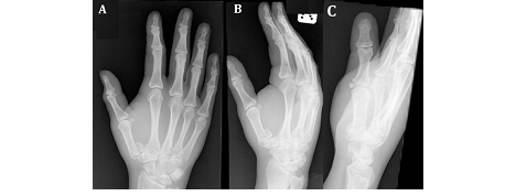
A 38-year old female presented to the emergency department with acute bruising and swelling of the right hand after falling down the stairs. On physical examination of the right hand, bony tenderness was noted in the first, second and third metacarpals and over first carpometacarpal joint. No open fracture was noted. Anteroposterior, oblique and lateral views of the right hand confirmed a fracture through the base of the first metacarpal with carpometacarpal joint disruption (Panel A-C). This type of fracture is also called Bennett fracture; this can occur following a fall, typically when an axial force is applied to a partially flexed first metacarpal bone.
As this was an unstable fracture, it was reduced with traction and consequently fixed with a K-wire. Post-fixation plain film demonstrated good joint positioning. The patient was then regularly seen in the fracture clinic, with follow-up plain films that demonstrated a good unification of the fracture. The patient then underwent a course of physiotherapy to regain full function of the right hand.

