A Rare and Aggressive Uterine Cancer: Carcinosarcoma
Article Information
Catarina Reis-de-Carvalho1*, André Borges2, Dusan Djokovic2
1Department of Obstetrics, Gynecology and Reproductive Medicine, Centro Hospitalar Universitário Lisboa Norte, Portugal
2Department of Obstetrics and Gynecology, Centro Hospitalar Lisboa Ocidental, Portugal
*Corresponding Author: Dr. Catarina Reis-de-Carvalho, Department of Obstetrics, Gynecology and Reproductive Medicine, Centro Hospitalar Universitário Lisboa Norte, Portugal
Received: 15 March 2020; Accepted: 02 April 2020; Published: 09 April 2020
Citation: Catarina Reis-de-Carvalho, André Borges, Dusan Djokovic. A Rare and Aggressive Uterine Cancer: Carcinosarcoma. Journal of Radiology and Clinical Imaging 3 (2020): 056-057.
Share at FacebookKeywords
Uterine carcinosarcoma; Mullerian tumor; Uterine cancer; Carcinosarcoma
Uterine carcinosarcoma articles, Mullerian tumor articles, Uterine cancer articles, Carcinosarcoma articles
Uterine carcinosarcoma articles Uterine carcinosarcoma Research articles Uterine carcinosarcoma review articles Uterine carcinosarcoma PubMed articles Uterine carcinosarcoma PubMed Central articles Uterine carcinosarcoma 2023 articles Uterine carcinosarcoma 2024 articles Uterine carcinosarcoma Scopus articles Uterine carcinosarcoma impact factor journals Uterine carcinosarcoma Scopus journals Uterine carcinosarcoma PubMed journals Uterine carcinosarcoma medical journals Uterine carcinosarcoma free journals Uterine carcinosarcoma best journals Uterine carcinosarcoma top journals Uterine carcinosarcoma free medical journals Uterine carcinosarcoma famous journals Uterine carcinosarcoma Google Scholar indexed journals Mullerian tumor articles Mullerian tumor Research articles Mullerian tumor review articles Mullerian tumor PubMed articles Mullerian tumor PubMed Central articles Mullerian tumor 2023 articles Mullerian tumor 2024 articles Mullerian tumor Scopus articles Mullerian tumor impact factor journals Mullerian tumor Scopus journals Mullerian tumor PubMed journals Mullerian tumor medical journals Mullerian tumor free journals Mullerian tumor best journals Mullerian tumor top journals Mullerian tumor free medical journals Mullerian tumor famous journals Mullerian tumor Google Scholar indexed journals Uterine cancer articles Uterine cancer Research articles Uterine cancer review articles Uterine cancer PubMed articles Uterine cancer PubMed Central articles Uterine cancer 2023 articles Uterine cancer 2024 articles Uterine cancer Scopus articles Uterine cancer impact factor journals Uterine cancer Scopus journals Uterine cancer PubMed journals Uterine cancer medical journals Uterine cancer free journals Uterine cancer best journals Uterine cancer top journals Uterine cancer free medical journals Uterine cancer famous journals Uterine cancer Google Scholar indexed journals Carcinosarcoma articles Carcinosarcoma Research articles Carcinosarcoma review articles Carcinosarcoma PubMed articles Carcinosarcoma PubMed Central articles Carcinosarcoma 2023 articles Carcinosarcoma 2024 articles Carcinosarcoma Scopus articles Carcinosarcoma impact factor journals Carcinosarcoma Scopus journals Carcinosarcoma PubMed journals Carcinosarcoma medical journals Carcinosarcoma free journals Carcinosarcoma best journals Carcinosarcoma top journals Carcinosarcoma free medical journals Carcinosarcoma famous journals Carcinosarcoma Google Scholar indexed journals Color Doppler score articles Color Doppler score Research articles Color Doppler score review articles Color Doppler score PubMed articles Color Doppler score PubMed Central articles Color Doppler score 2023 articles Color Doppler score 2024 articles Color Doppler score Scopus articles Color Doppler score impact factor journals Color Doppler score Scopus journals Color Doppler score PubMed journals Color Doppler score medical journals Color Doppler score free journals Color Doppler score best journals Color Doppler score top journals Color Doppler score free medical journals Color Doppler score famous journals Color Doppler score Google Scholar indexed journals irregular myometrial-endometrial junction articles irregular myometrial-endometrial junction Research articles irregular myometrial-endometrial junction review articles irregular myometrial-endometrial junction PubMed articles irregular myometrial-endometrial junction PubMed Central articles irregular myometrial-endometrial junction 2023 articles irregular myometrial-endometrial junction 2024 articles irregular myometrial-endometrial junction Scopus articles irregular myometrial-endometrial junction impact factor journals irregular myometrial-endometrial junction Scopus journals irregular myometrial-endometrial junction PubMed journals irregular myometrial-endometrial junction medical journals irregular myometrial-endometrial junction free journals irregular myometrial-endometrial junction best journals irregular myometrial-endometrial junction top journals irregular myometrial-endometrial junction free medical journals irregular myometrial-endometrial junction famous journals irregular myometrial-endometrial junction Google Scholar indexed journals Endometrial thickness articles Endometrial thickness Research articles Endometrial thickness review articles Endometrial thickness PubMed articles Endometrial thickness PubMed Central articles Endometrial thickness 2023 articles Endometrial thickness 2024 articles Endometrial thickness Scopus articles Endometrial thickness impact factor journals Endometrial thickness Scopus journals Endometrial thickness PubMed journals Endometrial thickness medical journals Endometrial thickness free journals Endometrial thickness best journals Endometrial thickness top journals Endometrial thickness free medical journals Endometrial thickness famous journals Endometrial thickness Google Scholar indexed journals endometrial canal articles endometrial canal Research articles endometrial canal review articles endometrial canal PubMed articles endometrial canal PubMed Central articles endometrial canal 2023 articles endometrial canal 2024 articles endometrial canal Scopus articles endometrial canal impact factor journals endometrial canal Scopus journals endometrial canal PubMed journals endometrial canal medical journals endometrial canal free journals endometrial canal best journals endometrial canal top journals endometrial canal free medical journals endometrial canal famous journals endometrial canal Google Scholar indexed journals uterine endometrial cancers articles uterine endometrial cancers Research articles uterine endometrial cancers review articles uterine endometrial cancers PubMed articles uterine endometrial cancers PubMed Central articles uterine endometrial cancers 2023 articles uterine endometrial cancers 2024 articles uterine endometrial cancers Scopus articles uterine endometrial cancers impact factor journals uterine endometrial cancers Scopus journals uterine endometrial cancers PubMed journals uterine endometrial cancers medical journals uterine endometrial cancers free journals uterine endometrial cancers best journals uterine endometrial cancers top journals uterine endometrial cancers free medical journals uterine endometrial cancers famous journals uterine endometrial cancers Google Scholar indexed journals multifocal vessel pattern articles multifocal vessel pattern Research articles multifocal vessel pattern review articles multifocal vessel pattern PubMed articles multifocal vessel pattern PubMed Central articles multifocal vessel pattern 2023 articles multifocal vessel pattern 2024 articles multifocal vessel pattern Scopus articles multifocal vessel pattern impact factor journals multifocal vessel pattern Scopus journals multifocal vessel pattern PubMed journals multifocal vessel pattern medical journals multifocal vessel pattern free journals multifocal vessel pattern best journals multifocal vessel pattern top journals multifocal vessel pattern free medical journals multifocal vessel pattern famous journals multifocal vessel pattern Google Scholar indexed journals
Article Details
1. Case Presentation
Uterine carcinosarcoma (UCS), previously known as malignant mixed Mullerian tumor, is a very rare tumor of the female reproductive tract, composed of both high-grade sarcomatous and carcinomatous elements [1]. In the United States its incidence is 1-4 per 100,000 women, is more frequently find in black women and the median age at diagnosis is between 62-67 years old [1]. Risk factors for developing UCS include advanced age, obesity, nulliparity, exogenous estrogen use, pelvic radiation, and tamoxifen [2]. Due to its aggressive nature, up to 60% have an extrauterine disease at presentation [3]. A pelvic ultrasound is the first-line imaging study. The typical appearance is a heterogeneous solid intrauterine mass with an irregular shape, hypoechoic and septated cystic echotexture, an ill-defined boundary, possible expansion of the endometrial canal, and abundant blood flow signals [4, 5]. UCS has a worse prognosis compared to other high-risk uterine endometrial cancers, with reported five-year disease-specific survival rates of 59% for stage I/II disease, 22% for stage III disease, and 9% for stage IV disease, regardless of therapy [2].
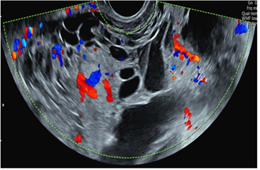
This image concerns a 58-year-old P1G1 woman with postmenopausal uterine bleeding and pelvic pain over the past 2 months. Ultrasound assessment, with endovaginal (Figure 1) and abdominal probes, revealed an enlarged uterus reaching the supraumbilical level and bearing a solid tumor with multiple cystic areas and a color Doppler score 4. The Pipelle sampling confirmed the diagnosis of carcinosarcoma in this patient who died two days later.

Figure 1: Ultrasound assessment, with endovaginal.
Transvaginal ultrasonography is appropriate for an initial evaluation of postmenopausal bleeding. Endometrial thickness of 4 mm, or less, has a greater than 99% negative predictive value for endometrial cancer [6]. The ultrasound is not accurate to distinguish carcinosarcomas from other endometrial malignancies since the features of high-grade endometrial cancer also include heterogeneous echogenicity, high Color Doppler score, multifocal vessel pattern, and irregular myometrial-endometrial junction [6]. However, symptoms and clinical signs, along with the recognition of suspected ultrasound features presented here, should trigger prompt medical assistance to improve patient survival.
References
- Ravishankar P, Smith DA, Avril S, et al. Uterine carcinosarcoma: a primer for radiologists. Abdominal Radiology 44 (2019): 2874-2885.
- Sagebiel TL, Bhosale PR, Patnana M, et al. Uterine Carcinosarcomas. Seminars in Ultrasound, CT and MRI. 40 (2019): 295-301.
- Denschlag D, Ulrich UA. Uterine carcinosarcomas - Diagnosis and management. Oncology Research and Treatment 41 (2018): 675-679.
- Zhao F, Xu Y, Zhang H, et al. Ultrasonographic findings of uterine carcinosarcoma. Gynecol Obstet Invest 84 (2019): 277-282.
- Hosh M, Antar S, Nazzal A, et al. Uterine Sarcoma: Analysis of 13,089 Cases Based on Surveillance, Epidemiology, and End Results Database. Int J Gynecol Cancer 26 (2016).
- Epstein E, Fischerova D, Valentin L, et al. Ultrasound characteristics of endometrial cancer as defined by International Endometrial Tumor Analysis (IETA) consensus nomenclature: prospective multicenter study. Ultrasound Obstet Gynecol 51 (2018): 818-828.
