Walker-Warburg Syndrome: A Case report of Congenital Muscular Dystrophy with Cerebro-Ocular Malformation, a Rare Syndrome in an African Patient
Article Information
Insiyah A Amiji1*, Maria N Bulimba1, Zawadi E Kalezi1, Janeth Fidelis Tarimo2, Karim P Manji1
1Department of Pediatrics and Child Health, Muhimbili University of Health and Allied Sciences, Dar es Salaam, Tanzania
2Muhimbili National Hospital, Dar es Salaam, Tanzania
*Corresponding Author: Insiyah A Amiji, Department of Pediatrics and Child Health Residency Program, Muhimbili University of Health and Allied Sciences, Dar es Salaam, Tanzania
Received: 03 June 2019; Accepted: 14 June 2019; Published: 24 June 2019
Citation:
Insiyah A Amiji, Maria N Bulimba, Zawadi E Kalezi, Janeth Fidelis Tarimo, Karim P Manji. Walker-Warburg Syndrome: A Case report of Congenital Muscular Dystrophy with Cerebro-Ocular Malformation, a Rare Syndrome in an African Patient. Journal of Pediatrics, Perinatology and Child Health 3 (2019): 069-075.
Share at FacebookAbstract
Walker-Warburg Syndrome is a genetically heterogeneous disease with autosomal recessive inheritance presenting with congenital muscular dystrophy with cerebro-ocular malformations since birth or infancy. Literature shows a mutation in Protein O-Mannosyltransferase 1 (POMT1) gene, which results in a defect in O-mannosylation of α-dystroglycan, these proteins are central to the pathogenesis of structural and functional brain abnormalities however, the exact pathophysiology of this disorder is not fully understood. The overall incidence still remains unknown and most cases have been reported from the Western hemisphere. In our setting we report a case of 3 weeks old male baby who was delivered by spontaneous vertex delivery at gestation age of 26 weeks with birth weight of 1900 gm referred to our hospital on the 6th day of life, with complaint of bilateral prominent, protruding eyes and inability to close the eyelids. The parents were non-consanguineous and this was the first child. No ante- natal ultrasound was done during the pregnancy. On further examination, he had macrocephaly 33 cm (>97th centile) with no fusion of the sutures, depressed nasal bridge, low set ears, bilateral dental lamina cyst, bilateral cortical thumb, micrognathia, and scoliosis. Systemic examination revealed a normal conscious level with weak cry, normal facial symmetry, generalized hypotonia and primitive reflex were weak to absent and ocular examination revealed bupthalmos, bilateral megalocornea and glaucoma. Creatine kinase was elevated-522 unit/L (0-25 units/L). Cranial Ultrasound revealed Dandy-Walker cyst in the posterior fossa cyst, with third ventricle dilatation (ventriculomegally), the corpus callosum was normal, Interventricular hemorrhage grade 2 and hyper echogenic eyeballs. Echocardiography revealed Double Outlet Right Ventricle. Management was supportive, aimed to improve growth through optimum feed
Keywords
Walker-Warburg Syndrome, Birth, Pregnancy, Children
Walker-Warburg Syndrome articles, Birth articles, Pregnancy articles, Children articles
Article Details
1. Introduction
Walker-Warburg Syndrome (WWS) is a rare form of a genetically heterogenous form of the disorder with autosomal recessive inheritance. It is characterized by congenital muscular dystrophy with oculo-cerebral malformations. It is the most severe form of congenital muscular dystrophy with most children dying before the age of three years. The most commonly mutated genes were discovered first including POMT1, POMT2, CRPPA, FKTN, FKRP and LARGE1 [1-2]. The overall incidence is unknown, but genetic studies done to evaluate various muscular dystrophies in Italy reports that the incidence of Dystroglycanopathies is 1.2 per 100,000 live births [3]. Symptoms and signs are already present at birth or in infancy, and are sometimes detected prenatally with imaging techniques. Clinical presentation includes variety of brain, eye and muscle abnormality like generalized hypotonia, muscle weakness, developmental delay with mental retardation and seizures in some children. Eye abnormalities include anterior eye anomalies (cataracts, shallow anterior chamber, microcornea and microphthalmia, and lens defects) and posterior eye anomalies (retinal detachment or dysplasia, hypoplasia or atrophy of the optic nerve and macula and coloboma). Glaucoma or buphthalmos may be present. Brain abnormalities include type II lissencephaly, hydrocephalus, ventriculomegally, vermal or general cerebellar hypoplasia and flat brainstem with small pyramids, hypoplasia/ agenesis of corpus callosum, occipital encephalocele and Dandy-Walker cystic malformation. Other features reported are genital abnormalities and facial dysmorphism such as low set or prominent ears and cleft lip or palate [1]. The overall prognosis is poor as it is the most severe form of congenital muscular dystrophy with most children dying before the age of three years.
2. Case Report
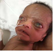
We report a case of 3 weeks old male baby who was delivered by spontaneous vertex delivery to primipara at the gestation age of 26 weeks, at Moro Hospital in Rufiji with a birth weight of 1900 gms. After birth, it was noted that there was bilateral prominent, protruding eyes and inability to close the eyelids. On the 6th day of life the baby was referred to the Muhimbili National Hospital. There was no reported history of seizures, eye discharge or fevers. The parents were not consanguineous and the mother had 4 miscarriages before this pregnancy, which were in early trimester. She made four antenatal care visits received anti-malarial, hematinic and tetanus vaccination, HIV status and syphilis status was negative, however, she had a urinary tract infection one week prior to delivery that was treated. No ante-natal ultrasound was done throughout the pregnancy. On general examination, we found macrocephaly 33 cm (>97th centile) but no fusion of the sutures, depressed nasal bridge, low set ears, bilateral dental lamina cyst, bilateral cortical thumb, micrognathia, and scoliosis. Neurologic examination revealed normal conscious level, weak cry, baby normal facial symmetry, no tongue deviation or fasciculations, cranial nerves II, III, IV, VI, IX, XI were difficult to assess. Baby had generalized hypotonia and primitive reflexes were weak to absent. Ocular examination revealed bupthalmos, bilateral megalocornea and glaucoma.
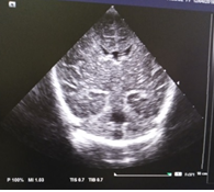
CBC were within normal ranges, Creatinine 17.7 mmol/l, Urea 4.4 mmol/L, Sodium 137 mmol/l, Potassium 4.4 mmol/l, Calcium 2.56 mmol/l. Serum Creatine kinase was elevated- 522 unit/L (0-25 units/L). TORCHES screening was positive for Rubella IgG, but negative for CMV, rubella IgM, toxoplasmosis. Cranial Ultrasound revealed Dandy-Walker cyst in the posterior fossa cyst, with third ventricle dilatation (ventriculomegally), the corpus callosum was normal, Interventricular hemorrhage grade 2 and hyper echogenic eyeballs. ECHO was done which revealed Double Outlet Right Ventricle and the antifailures were initiated. Tarsorraphy was planned, but due to the underlying heart condition the procedure was postponed until when he was stabilized. The baby was planned for Tarsorraphy but due to the underlying heart condition the procedure was postponed until when he was stabilized. Genetic testing was not done as it is not available in our setting.
Management was supportive. During the course of hospitalization the baby was treated with Ciprofloxacin eye drops for exposure keratitis, eye padding and diuretics (syrup furosemide 2 mg once daily) for the underlying heart condition, antibiotics for pneumonia and expressed breast feeding. After one month the baby had gained 200 gms and weighed 2100 gm. On the 30th day of life baby aspirated milk contents while feeding and developed aspiration pneumonia and was kept on Continuous positive airway pressure (CPAP). Unfortunately, the baby died on the 42nd day of life due to respiratory failure. The overall prognosis was poor due to multisystemic congenital abnormalities. Mother was counselled from the beginning and she was cooperative throughout the course of illness. Genetic counseling was done to the mother. She was also counselled to plan the next pregnancy and ensure that ante- natal ultrasound should be done earlier in the pregnancy as brain abnormalities has been detected as early as 13 weeks. Dandy Walker cysts in the posterior fossa, venticulomegally, macrocephaly, generalized hypotonia, eye abnormalities and raised CK and the cranial USS characteristics described above alerts to the possibility of WWS.
3. Discussion
WWS is a rare syndrome and has a spectrum of various eye, brain and muscle abnormalities. It carries a poor prognosis. Eye malformations include microphthalmia, coloboma, glaucoma, cataract, megalocornea, persistent hyperplastic primary vitreous and retinal dysplasia or detachment. The ophthalmological features were no less variable and the abnormalities of the anterior segment seem to be as frequent, in our case being megalocornea and congenital glaucoma that could be suggestive of retinal detachment and are mentioned in all the reports. CNS malformations include absent laminar structure agyria/pachygyria, ventriculomegaly, cerebellar dysplasia, encephalocele, Dandy-Walker cyst, hydrocephalus [4-7]. Despite of many cases of congenital muscular dystrophies in African countries, there is no report of WWS. Most cases are reported in the literature are from the Western hemisphere. Laboratory investigations usually show elevated creatine kinase, myopathic/dystrophic muscle pathology and altered α-dystroglycan.
Familial recurrence of this syndrome has been reported in consanguineous parents, which strongly suggests autosomal recessive inheritance, [8] In our case the parents were non consanguinoues and various cases have also been reported in this population [9-10]. Agarwal, et al. [11] reported a similar case with dandy walker cysts in the posterior horn and eye abnormalities such as micropthalmos and megalopthamos in the same patient. Pagon R, et al. [6] has also reported case of 2 siblings with eye and brain abnormality where one had a progressive hydrocephalus and central corneal opacities also known as Peters anomaly and a 2 cm skin covered encephalocele, hydrocephalus and micropthalmia of the right eye and megalocornea of the left eye and retinal detachment in the other one. The infants, however, died at one year of age, with increasing spasticity. Heyer R, et al. [12], Pavone L, et al. [9] have also described congenital muscular dystrophy characterized by muscular weakness, hypotonia and joint contractures to be associated with eye and cerebral malformations in Walker Warburg syndrome. In addition to the congenital malformations, elevated Creatinine Kinase values were reported in all of the above cases and this was also observed in our baby.
|
|
CASE REPORT |
Our case 2019 |
Heyer et al. [12] |
Murphy et al. [13] |
Fawzi et al. [14] |
S. Agarwal et al. [15] |
Agarwal et al. [11] |
|
Gender |
Male |
Male |
Female |
Male |
Male |
Female |
|
|
CNS malformation |
|||||||
|
Agyria/pachygyria |
- |
- |
- |
- |
- |
||
|
Cerebral atrophy |
+ |
+ |
- |
- |
- |
- |
|
|
Cerebellar dysplasia |
- |
- |
- |
+ |
- |
||
|
Dandy walker cyst |
+ |
+ |
- |
- |
- |
+ |
|
|
Hydrocephalus |
- |
+ |
- |
+ |
+ |
||
|
Ventriculomegally |
+ |
- |
- |
- |
+ |
+ |
|
|
Encephalocele |
- |
- |
- |
- |
- |
- |
|
|
OFC (cm) |
33 cm |
- |
34.5 |
34 |
- |
- |
|
|
Lissencephaly |
- |
- |
- |
- |
+ |
- |
|
|
Ocular malformation |
|||||||
|
Retinal dysplasia/ detachment |
+ |
- |
Left + |
- |
- |
||
|
Corneal opacity |
+ |
- |
+ |
Right + |
- |
- |
|
|
Micropthalmia |
- |
- |
- |
- |
+ |
Right |
|
|
Megalopthalmia |
+ |
- |
- |
- |
- |
left |
|
|
Megalocornea |
- |
- |
- |
+ |
- |
- |
|
|
Glaucoma |
+ |
+ |
- |
+ |
- |
- |
|
|
Cataract |
- |
- |
- |
- |
- |
- |
|
|
Intraocular pressure |
- |
- |
increased |
- |
- |
Increased |
|
|
Muscular abnormality |
|||||||
|
Hyptonia |
+ |
+ |
+ |
+ |
+ |
+ |
|
|
Reduced/ weak Deep tendon reflex |
+ |
+ |
+ |
+ |
- |
- |
|
|
Contractures |
- |
+ |
- |
- |
- |
+ |
|
|
Scoliosis |
+ |
+ |
- |
- |
- |
- |
|
|
Hip dislocation |
- |
- |
- |
- |
- |
+ |
|
|
Creatinine kinase levels |
Raised |
Raised |
Raised |
Raised |
Raised |
Raised |
|
|
Others Facial dysmorphism |
+ |
- |
+ |
+ |
- |
+ |
|
Table 1: Comparison of present case with different cases.
Management is usually supportive and preventive. Genetic counselling should be done as it is an autosomal recessive disorder and in families with one affected child, the risk of having another child with the disease is 25%. Increased awareness of this rare but lethal condition, and a high index of suspicion during a routine antenatal ultrasound could prompt further advanced fetal ultrasonography and magnetic resonance imaging, and aid in timely prenatal diagnosis, management, and counseling.
References
- Vajsar J, Schachter H. Walker-Warburg syndrome. Orphanet J Rare Dis 1 (2006): 1-5.
- Reeuwijk J Van, Janssen M, Elzen C Van Den, et al. POMT2 mutations cause alpha-dystroglycan hypoglycosylation and Walker Warburg syndrome. J Med Genet 42 (2005): 907-912.
- Graziano A, Messina S, Bruno C, et al. Prevalence of congenital muscular dystrophy in Italy A population study. Am Acad Nerology 84 (2015): 904-911.
- Walker AE. Lissencephaly. Arch NeurPsych 48 (1942): 13-29.
- Warburg M. Heterogeneity of Congenital Retinal Non-Attachment, Falciform Folds and Retinal Dysplasia. Hum Hered 26 (1976): 137-148.
- Pagon RA, Clarren SK, Milam DF, et al. Autosomal recessive eye and brain anomalies. Warburg syndrome. J Pediatr 4 (1983): 542-546.
- Garner RMW. Hydrocephalus, agyria, pseudoencephalocele, retinal dysplasia, and anterior chamber anomalies. J Med Genet 18 (1981): 314-318.
- Ayme S, Mattei J. HARD (E) Syndrome: Report of a Sixth Family With Support for Autosomal-Recessive Inheritance 766 (1983): 759-766.
- Pavone L, Gullotta F, Grasso S, et al. Hydrocephalus, Lissencephaly, Ocular Abnormalities and Congenital Muscular Dystrophy. A Warburg Syndrome Variant? Neuropediatrics 17 (1986): 206-211.
- Bordarier C, Aicardi J, Goutieres F. Congenital Hydrocephalus and Eye Abnormdties with Severe Developmental Brain Defects: Warburg’ s Syndrome. Ann Neurol 16 (1984): 60-65.
- Agrawal P, Patel C. Walker Warburg syndrome associated with microphthalmos and megalophthalmos: A rare combination. Oman J Opthalmology 6 (2013): 5-7.
- Heyer R, Ehrich J, Goebel H. Congenital Muscular Dystrophy with Cerebral and Ocular Malformations (Cerebro-Oculo-Muscular Syndrome). Brain Dev 8 (1986): 614-619.
- Murphy KJ, Pebenito R, Storm RL, et al. Walker-Warburg syndrome Case report and literature review Ophthalmic Paediatr Genet 11 (1990): 103-108.
- Fawzi M, Bessisso M, Omar F. CASE REPORT WALKER-WARBURG SYNDROME: A Case Report of a Qatari Patient. Qatar Med J 9 (2000): 66-67.
- Agrawal S. Walker-Warburg syndrome. Br Med J (2011): 3-4.
- Low ASC, Lee SL, Tan ASA, et al. Difficulties with Prenatal Diagnosis of the Walker- Warburg Syndrome Difficulties with Prenatal Diagnosis of the Walker-Warburg Syndrome. Acta radiol 46 (2009): 645-651.


