Laparoscopic Cholecystectomy for a giant Gallstone
Article Information
Irfan Jan Khan1*, Murad Alahmad2, Fahad Aurif3, Najeeb Hussain Mir4
1Associate Consultant General Surgery HMC, Qatar
2Resident General Surgery HMC, Qatar
3Specialist General Surgery HMC, Qatar
4Consultant General Surgery HMC, Qatar
*Corresponding Author: Irfan Jan Khan, Associate Consultant General Surgery HMC, Qatar.
Received: 02 December 2021; Accepted: 22 December 2021; Published: 25 January 2022
Citation: Irfan Jan Khan, Murad Alahmad, Fahad Aurif, Najeeb Hussain Mir. Laparoscopic Cholecystectomy for a giant Gallstone. Archives of Clinical and Medical Case Reports 6 (2022): 40-44.
Share at FacebookAbstract
Introduction: Cholelithiasis is very common, but giant gallstones - gallstones bigger than 5 cm in diameter, are very rare. It is very challenging to remove them laparoscopically, especially in emergency situations.
Case Report: A 69-year-old female complained of right upper abdominal pain with vomiting for 1 day. Abdominal ultrasound indicated acute cholecystitis and a single, extremely large gallstone (9cm). Emergency laparoscopic cholecystectomy was performed successfully.
Discussion: Gallstone over 5 cm in diameter is very rare. Laparoscopic Cholecystectomy (LC) is very difficult for these cases, especially in the emergency situations. Laparoscopic cholecystectomy can, however, be safely performed in experienced hands. To the best of our knowledge, this is one of the biggest gallstones managed laparoscopically.
Conclusion: Although the presence of giant gallstones poses technical challenges, laparoscopic cholecystectomy can be safely performed in the experienced hands. However, if the anatomy cannot be delineated clearly, open surgery remains the safe option.
Keywords
Cholelithiasis; Giant gallstones; Laparoscopic cholecystectomy
Article Details
1. Introduction
Gallstone disease is one of the commonest surgical problems with women being affected more common than men (1). Laparoscopy has become the gold standard for the treatment of symptomatic gallstones. Gallstones with a diameter of over 5 cm are very rare. Such cases are difficult for LC, especially in the emergency situations. We present a case of a 9 cm giant gallstone which was removed laparoscopically.
2. Case Report
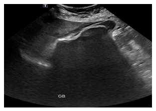
A 69-year-old diabetic, hypertensive female known to have WPW syndrome (status post ablation) with past history of DVT and CVA presented to the emergency with one-day history of upper abdominal pain associated with vomiting. There was no h/o fever. Clinical examination revealed a tender right hypochondrium with a positive Murphy’s sign. Laboratory tests revealed leukocytosis (21000) and high CRP (256). Ultrasound abdomen was suggestive of acute cholecystitis with a single large gallstone (Figure 1). Her present medications included Aspirin in addition to antihypertensives and antidiabetics. After cardiac evaluation, she was posted for emergency surgery. Laparoscopic cholecystectomy was performed and patient had an uneventful recovery.
3. Discussion
Gallstone disease is one of the commonest surgical problems with women being affected more common than men [1]. Laparoscopy has become the gold standard for the treatment of symptomatic gallstones and can be performed in 96% of the cases. The rate of conversion from laparoscopic to open cholecystectomy is about 4–5% [2]. Gallstones with a diameter of over 5 cm are very rare. Such cases are difficult for LC, especially in the emergency situations. Some surgeons may consider a giant gallstone an indication for open surgery [3]. The risk of conversion is related to surgeon factors, patient factors, and possibly equipment factors [4]. We present a case of a 9 cm giant gallstone which was removed laparoscopically.
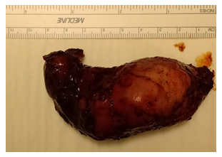
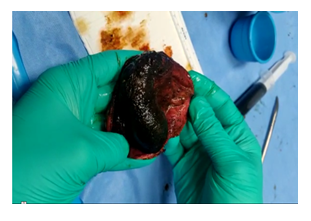
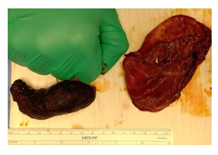
The operation was performed using 4 ports with standard port placement. Because of presence of a large gallstone, gallbladder could not be grasped in the usual fashion. Aspiration of about 10-20ml of thick bile allowed the fundus to be grasped and retracted towards the right shoulder. The very presence of a giant gallstone did not allow a clear view of Hepatocystic triangle. After some dissection in this area, it was decided to dissect the gallbladder off the liver bed retrogradely. The presence of edema helped in getting into the tissue planes relatively easily. During all the dissection, it was however difficult to grasp the gallbladder with laparoscopic instruments. The manipulation of gallbladder was carried out using opened jaws of a bowel grasper, the so called “Fan technique” [5]. Cystic duct and Cystic artery were clearly identified, clipped and cut. Gallbladder was retrieved in an enobag through the camera port. The camera port needed to be extended to retrieve the specimen (Figure 2). The patient had an uneventful recovery and was discharged on 3rd postoperative day. A 9 cm gallstone was found within this huge gallbladder (Figure 3). Singh Y el al have reported a successful retrieval of a 13cm gallstone laparoscopically [6]. Xu et al. reported the laparoscopic retrieval of a 9.5-cm gallstone [7], and Becerra et al. reported removal of a 16.8-cm long gallstone via classical cholecystectomy in the emergency setting [8]. From the literature to date, our gallstone appears to be one of the largest removed laparoscopically. Giant gallstones can pose difficulties in two different ways. First, larger stones can result in a thicker gallbladder wall secondary to inflammation. The thickness of the gallbladder wall has been shown to correlate with conversion to open surgery in bivariate analyses [9]. Second, a large gallstone can lead to technical difficulties, for example, grasping the gallbladder with laparoscopic instruments may be extremely difficult and also to gain appropriate anatomical exposure for safe dissection.
4. Conclusion
Although the presence of giant gallstones poses technical challenges, laparoscopy can be safely performed in experienced hands. However, if the anatomy cannot be delineated clearly, open surgery remains the safe option.
References
- Lammert F, Sauerbruch T. Mechanisms of disease: the genetic epidemiology of gallbladder stones. Nat Clin Pract Gastroenterol Hepatol 2 (2005): 423-433.
- Berthou JCh, Dron B, Charbonneau P, et al. Evaluation of laparoscopic treatment of common bile duct stones in a prospective series of 505 patients: indications and results. Surg Endosc 21 (2007): 1970-1974.
- Dalal S, Pankaj, Bohriwal S, et al. Giant Gallstone: A rare indication of open cholecystectomy. JCR 4 (2014):17-19.
- Kama NA, Doganay M, Dolapci M, et al. Risk factors resulting in conversion of laparoscopic cholecystectomy to open surgery. Surg Endosc 15 (2001): 965-968.
- Litwin DEM, Cahan MA. Laparoscopic Cholecystectomy. Surgical Clinics of North America 88 (2008): 1295-1313.
- Singh Y, Mohammed S, Hosein A, Ramoutar K, Narayansingh V A Giant Gallstone: The Largest Gallstone Removed Laparoscopically in the World. Cureus 12 (2020): 7933.
- Xu X, Hong T, Zheng C. Giant gallstone performed by emergency laparoscopic cholecystectomy. Int J Surg Case Rep 4 (2013): 1163-1164.
- Becerra P, Becerra V, Aguilar C, et al. Giant gallstone: a case report. Int J Surg Case Rep 2 (2011): 228-229.
- Raman SR, Moradi D, Samaan BM, et al.: The degree of gallbladder wall thickness and its impact on outcomes after laparoscopic cholecystectomy. Surg Endosc 26 (2012): 3174-3179.


