Calcific Tendinitis of the Patellar Tendon with MRI Correlations
Article Information
Nicholas Newcomb BS1*, Bhargav Venkatraghavan BS2, Paras Sahani BS2, Eric Lin BA3
Heather Yerdon BS1, Piers Barry MD4
1University of Queensland-Ochsner Clinical School, New Orleans, Louisiana, USA
2University of California-San Diego, San Diego, California, USA
3University of California-Berkeley, Berkeley, California, USA
4Piers Barry MD Inc, San Francisco, California, USA
*Corresponding Author: Nicholas Newcomb, University of Queensland-Ochsner Clinical School, 514 Jefferson Hwy. New Orleans, LA USA
Received: 30 June 2021; Accepted: 12 July 2021; Published: 31 July 2021
Citation: Nicholas Newcomb BS, Bhargav Venkatraghavan BS, Paras Sahani BS, Eric Lin BA, Heather Yerdon BS, Piers Barry MD. Calcific Tendinitis of the Patellar Tendon with MRI Correlations. Journal of Orthopaedics and Sports Medicine 3 (2021): 116-122.
Share at FacebookAbstract
Background: Calcific tendinitis is a predominantly cell mediated disease that has been well documented in multiple areas of the body, but very rarely in the patellar tendon. This study presents an unusual case of severe patellar calcific tendinitis initially diagnosed as mucoid degeneration and partial patellar tendon tear after a sports injury. Three modalities of diagnostic imaging were unable to identify calcific deposition which was only discoverable intra-operatively. We describe this unusual presentation and optimal imaging sequences for diagnosis.
Case Report: A 51-year-old male presented three weeks after injuring his left knee playing tennis. The patient lunged forward with his left foot to hit a ball when he felt a sudden sharp pulling sensation at his anterior knee over the patellar tendon. This pain was most pronounced with weight-bearing activities and did not improve over the next month. Left knee radiographs were unremarkable; MRI and ultrasound images demonstrated diffuse patellar tendon partial thickness interstitial tearing and what appeared to be mucoid degeneration. None of the standard imaging modalities identified calcification. Surgical exploration of the patellar tendon revealed a large pocket of classic calcific deposition under pressure which was evacuated prior to tendon repair. The patient had rapid resolution of his pain and was full weight bearing at two weeks post-operation.
Conclusion: Calcific tendinitis of the patellar tendon is an unusual condition that can present diagnostic difficulty. Commonly used imaging modalities and protocols may be insufficient in diagnosing calcification. If tendon calcification is suspected, consideration may be given to the use of gradient-echo sequences as a part of MRI imaging for definitive diagnosis and treatment optimization.
Keywords
Patellar tendinitis; Calcific tendinitis; Patellar tendon rupture; Tendon repair
Calcific tendinitis articles Calcific tendinitis Research articles Calcific tendinitis review articles Calcific tendinitis PubMed articles Calcific tendinitis PubMed Central articles Calcific tendinitis 2023 articles Calcific tendinitis 2024 articles Calcific tendinitis Scopus articles Calcific tendinitis impact factor journals Calcific tendinitis Scopus journals Calcific tendinitis PubMed journals Calcific tendinitis medical journals Calcific tendinitis free journals Calcific tendinitis best journals Calcific tendinitis top journals Calcific tendinitis free medical journals Calcific tendinitis famous journals Calcific tendinitis Google Scholar indexed journals Shoulder articles Shoulder Research articles Shoulder review articles Shoulder PubMed articles Shoulder PubMed Central articles Shoulder 2023 articles Shoulder 2024 articles Shoulder Scopus articles Shoulder impact factor journals Shoulder Scopus journals Shoulder PubMed journals Shoulder medical journals Shoulder free journals Shoulder best journals Shoulder top journals Shoulder free medical journals Shoulder famous journals Shoulder Google Scholar indexed journals Wrist articles Wrist Research articles Wrist review articles Wrist PubMed articles Wrist PubMed Central articles Wrist 2023 articles Wrist 2024 articles Wrist Scopus articles Wrist impact factor journals Wrist Scopus journals Wrist PubMed journals Wrist medical journals Wrist free journals Wrist best journals Wrist top journals Wrist free medical journals Wrist famous journals Wrist Google Scholar indexed journals Hip articles Hip Research articles Hip review articles Hip PubMed articles Hip PubMed Central articles Hip 2023 articles Hip 2024 articles Hip Scopus articles Hip impact factor journals Hip Scopus journals Hip PubMed journals Hip medical journals Hip free journals Hip best journals Hip top journals Hip free medical journals Hip famous journals Hip Google Scholar indexed journals Thigh articles Thigh Research articles Thigh review articles Thigh PubMed articles Thigh PubMed Central articles Thigh 2023 articles Thigh 2024 articles Thigh Scopus articles Thigh impact factor journals Thigh Scopus journals Thigh PubMed journals Thigh medical journals Thigh free journals Thigh best journals Thigh top journals Thigh free medical journals Thigh famous journals Thigh Google Scholar indexed journals Knee articles Knee Research articles Knee review articles Knee PubMed articles Knee PubMed Central articles Knee 2023 articles Knee 2024 articles Knee Scopus articles Knee impact factor journals Knee Scopus journals Knee PubMed journals Knee medical journals Knee free journals Knee best journals Knee top journals Knee free medical journals Knee famous journals Knee Google Scholar indexed journals Ankle articles Ankle Research articles Ankle review articles Ankle PubMed articles Ankle PubMed Central articles Ankle 2023 articles Ankle 2024 articles Ankle Scopus articles Ankle impact factor journals Ankle Scopus journals Ankle PubMed journals Ankle medical journals Ankle free journals Ankle best journals Ankle top journals Ankle free medical journals Ankle famous journals Ankle Google Scholar indexed journals Foot articles Foot Research articles Foot review articles Foot PubMed articles Foot PubMed Central articles Foot 2023 articles Foot 2024 articles Foot Scopus articles Foot impact factor journals Foot Scopus journals Foot PubMed journals Foot medical journals Foot free journals Foot best journals Foot top journals Foot free medical journals Foot famous journals Foot Google Scholar indexed journals Sports injury articles Sports injury Research articles Sports injury review articles Sports injury PubMed articles Sports injury PubMed Central articles Sports injury 2023 articles Sports injury 2024 articles Sports injury Scopus articles Sports injury impact factor journals Sports injury Scopus journals Sports injury PubMed journals Sports injury medical journals Sports injury free journals Sports injury best journals Sports injury top journals Sports injury free medical journals Sports injury famous journals Sports injury Google Scholar indexed journals Knee injury articles Knee injury Research articles Knee injury review articles Knee injury PubMed articles Knee injury PubMed Central articles Knee injury 2023 articles Knee injury 2024 articles Knee injury Scopus articles Knee injury impact factor journals Knee injury Scopus journals Knee injury PubMed journals Knee injury medical journals Knee injury free journals Knee injury best journals Knee injury top journals Knee injury free medical journals Knee injury famous journals Knee injury Google Scholar indexed journals Playing tennis articles Playing tennis Research articles Playing tennis review articles Playing tennis PubMed articles Playing tennis PubMed Central articles Playing tennis 2023 articles Playing tennis 2024 articles Playing tennis Scopus articles Playing tennis impact factor journals Playing tennis Scopus journals Playing tennis PubMed journals Playing tennis medical journals Playing tennis free journals Playing tennis best journals Playing tennis top journals Playing tennis free medical journals Playing tennis famous journals Playing tennis Google Scholar indexed journals Osteophytes articles Osteophytes Research articles Osteophytes review articles Osteophytes PubMed articles Osteophytes PubMed Central articles Osteophytes 2023 articles Osteophytes 2024 articles Osteophytes Scopus articles Osteophytes impact factor journals Osteophytes Scopus journals Osteophytes PubMed journals Osteophytes medical journals Osteophytes free journals Osteophytes best journals Osteophytes top journals Osteophytes free medical journals Osteophytes famous journals Osteophytes Google Scholar indexed journals Patellar tendon articles Patellar tendon Research articles Patellar tendon review articles Patellar tendon PubMed articles Patellar tendon PubMed Central articles Patellar tendon 2023 articles Patellar tendon 2024 articles Patellar tendon Scopus articles Patellar tendon impact factor journals Patellar tendon Scopus journals Patellar tendon PubMed journals Patellar tendon medical journals Patellar tendon free journals Patellar tendon best journals Patellar tendon top journals Patellar tendon free medical journals Patellar tendon famous journals Patellar tendon Google Scholar indexed journals Arthroscopy articles Arthroscopy Research articles Arthroscopy review articles Arthroscopy PubMed articles Arthroscopy PubMed Central articles Arthroscopy 2023 articles Arthroscopy 2024 articles Arthroscopy Scopus articles Arthroscopy impact factor journals Arthroscopy Scopus journals Arthroscopy PubMed journals Arthroscopy medical journals Arthroscopy free journals Arthroscopy best journals Arthroscopy top journals Arthroscopy free medical journals Arthroscopy famous journals Arthroscopy Google Scholar indexed journals Chondrocytes articles Chondrocytes Research articles Chondrocytes review articles Chondrocytes PubMed articles Chondrocytes PubMed Central articles Chondrocytes 2023 articles Chondrocytes 2024 articles Chondrocytes Scopus articles Chondrocytes impact factor journals Chondrocytes Scopus journals Chondrocytes PubMed journals Chondrocytes medical journals Chondrocytes free journals Chondrocytes best journals Chondrocytes top journals Chondrocytes free medical journals Chondrocytes famous journals Chondrocytes Google Scholar indexed journals
Article Details
1. Introduction
Calcific tendinitis is a cell mediated disease, characterized by the metaplastic transformation of tenocytes into chondrocytes, resulting in calcific deposits in tendons [1]. The progression of calcific tendinitis through a pre-calcific phase, calcification phase, stable phase, and resorptive phase has been well documented. Initially, vulnerable sites undergo cellular changes predisposing them to calcification in the pre-calcific phase.
This is followed by cellular secretion of chalky deposits in the calcification phase and a subsequent painless stable phase. The final resorptive phase is most painful and may last for weeks while the calcium deposit develops a toothpaste-like appearance [2]. Clinically, patients with calcific tendinitis may present with acute onset or chronic pain and restricted range of motion [3].
Calcific tendinitis is most commonly seen in the rotator cuff tendons of the shoulder; their incidence is as high as 2.7%, with approximately 35% occurring asymptomatically [3]. Calcific tendinitis has also been documented at the wrist, hip, thigh, knee, ankle, and foot [3]. Very few cases of calcific tendinitis of the patellar tendon have been documented. Calcification can be visualized on radiographic, ultrasound, and MR imaging but is typically non-specific. On radiographic imaging, calcific deposits may appear well defined (in the calcification phase) or translucent (in the resorptive phase). On ultrasound examination of the shoulder, these deposits usually appear hyperechoic in the calcification phase or isoechoic in the resorptive phase. Calcifications appear as vague regions of low signal on T1 and T2 MR imaging [4]. Tendon calcification can often be missed on standard imaging, and in very rare cases, may mimic tendon rupture [5].
We present here a rare case of severe patellar calcific tendonitis initially diagnosed as mucoid degeneration and partial tear of the patellar tendon after a sports injury. Three modalities of diagnostic imaging including ultrasound, radiographs, and MRI were unable to correctly diagnose calcific deposition. The diagnosis was made after surgical exploration.
2. Case Report
A 51-year-old male with a BMI of 32 presented three weeks after injuring his left knee while playing tennis. The patient described pushing off with his left foot as he lunged forward for a ball and felt a sudden sharp pulling sensation at his anterior knee. Immediately following the injury, he felt soreness and pain and developed left knee swelling that night. Oral non-steroidal anti-inflammatories (NSAIDs) and ice helped minimally. He continued to have sharp anterior knee pain, inferior to the patella for multiple weeks. His pain worsened when weight-bearing, walking, or climbing stairs.
On examination, he had a mildly antalgic gait. He had mild effusion and popliteal cyst which had improved since his injury. Passive knee extension was full to 0 degrees without discomfort and flexion was 135 degrees with discomfort at the anterior aspect of the knee. The patient was tender to palpation over the patellar tendon. There was no tenderness to palpation along the medial or lateral joint lines. McMurray’s test was normal (tests for meniscal injury). The patient had 1+ translation with both Lachman and Anterior Drawer tests, a normal Posterior Drawer test and was stable to both Varus and Valgus stress (respective tests for anterior, posterior, lateral, and medial ligamentous injuries). The patella was stable without apprehension and painless with the patellar grind test (evaluates for patellar chondral injury).
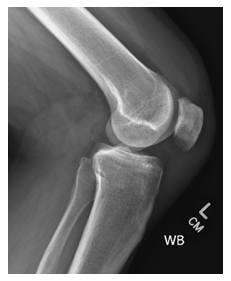
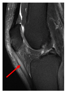
The patient completed left knee 4-view radiographs 8 days post injury (Image 1). There was no indication of fracture or dislocation. A diagnostic ultrasound was performed which showed partial tearing of the patellar tendon with small, marginal osteophytes at the inferior aspect of the patella. The patient completed a six sequence, closed, 3 Tesla MRI 21 days post-injury (Image 2).
Gradient echo sequences were not performed. An interstitial tear of the patellar tendon was visualized. There was a thick ‘mucoid’ component 3.4 cm superior-to-inferior and 5 mm in diameter. There was mild fraying at the free edge body of the lateral meniscus.

Image 1: Left knee radiograph, lateral view. Bony structures, alignment, and joint spaces are unremarkable.

Image 2:Left knee MRI, 3 tesla, PD fat suppressed, sagittal cut. The arrow demonstrates what appears to be prominent mucoid component seen at the patellar tendon, 3.4 cm superior-to-inferior and 5 mm in diameter.

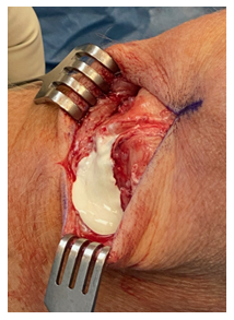
Image 3: Intraoperative image of anterior, vertical incision of the left knee. A large calcific deposit is seen draining from within the patellar tendon.
Surgical intervention was performed 45 days after initial complaint to explore and repair the patellar tendon tear after the patient had not improved with rest, activity modification, bracing, cryotherapy, or NSAIDs. Surgery included open patellar tendon anterior-posterior repair with removal of the calcific deposits and arthroscopic meniscal debridement. At arthroscopy, calcific deposits were seen and removed from the knee joint anterior to the ACL. A posterior horn tear of the medial meniscus and anterior horn tear of the lateral meniscus were debrided. The patellofemoral compartment and remaining cartilage in the knee were in good condition. A 10 cm midline incision was made, and a large inflamed prepatellar bursa was found along with thickening of the patellar tendon. Upon opening the patellar paratenon, there was a longitudinal split in the lateral side and thickening in the medial side of the tendon. The patellar tendon had calcific deposits under pressure which expelled from the tendon when opened (Image 3).
Degeneration and partial thickness tearing were found in the tendon. The tendon was debrided, and the calcific deposits were removed. The anterior and posterior layers of the tendon were sutured together. The longitudinal tear in the tendon was repaired using side-to side-suturing.
The patient was allowed to weight-bear as tolerated two weeks after surgery. Physical Therapy facilitated rapid return to full range of motion. By 8 weeks post-surgery, he was able to walk 5 miles and ride a stationary bike without limitations. His calcific deposits have not recurred.
3. Discussion
Calcific tendonitis of the patellar tendon is an uncommon condition that has not been well documented thus far [5, 6]. X-ray, MRI, and ultrasound are often used in evaluating knee injuries, however these imaging modalities and protocols may be insufficient in diagnosing calcific tendonitis. Calcific tendonitis is typically seen as a loss in signal intensity on standard MRI sequences, however these sequences pose diagnostic difficulty due to the low number of resonating protons in calcium deposits [4]. As a result, alternative MRI imaging techniques such as susceptibility weighted imaging (SWI) and gradient echo sequences (GREs), have been considered [7]. Typical MRI sequences consist of spin echo sequences such as T1-weighted sequences and T2-weighted sequences in which tendons are visualized as low signal intensities. GREs, however, visualize tendons with high signal intensities due to shorter echo times in comparison to spin echo sequences [8].
GREs are particularly useful in detecting calcific tendonitis as these sequences reveal positive phase shifts which are absent on standard MR imaging [9]. As a result, these imaging techniques can be used to detect calcific tendonitis which is not seen on x-ray, ultrasound, and standard MRI. However, GREs have been shown to be susceptible to signal artifacts [10]. The presence of air/tissue boundaries or metal implants limit the diagnostic ability of GREs due to the presence of void signals and susceptibility artifacts. These challenges have resulted in limited use of GREs for imaging tendons despite their increased sensitivity for calcium deposition.
The typical treatment for calcific tendinitis consists of physical therapy, NSAIDs, cortisone injections, or surgery [11]. Surgery is typically recommended if the patient fails conservative management or if there is a significant tendon tear. Standard surgical procedures include calcific deposit removal, bursectomy, and tendon repair [12].
In our case, the patient failed conservative treatment including bracing, cryotherapy, and oral NSAIDs and had pain and difficulty with activities of daily living for six weeks. The patient underwent surgical intervention with a successful outcome.
4. Conclusion
Calcific tendinitis of the patellar tendon is an unusual condition that can present diagnostic difficulty. Commonly used imaging modalities and protocols may be insufficient in diagnosing calcification. Although rare, patellar tendon calcific tendonitis may be better visualized with GRE MRI sequences for improved preoperative understanding of treatment and expected outcomes.
Disclosures
The authors have no financial disclosures or conflicts of interest to declare.
Ethical Approval and Consent
Institutional review board approval is not required for a deidentified single case report. Informed consent was obtained from the patient for the publication of this case report.
References
- De Carli, A et al. Calcific tendinitis of the shoulder. Joints 2, 3 (2014): 130-136.
- Uhthoff, Hans K. et al. Calcific tendinopathy of the rotator cuff: pathogenesis, diagnosis, and management. Journal of the American Academy of Orthopaedic Surgeons 5, 4 (1997): 183-191.
- Holt PD, Keats TE. Calcific tendinitis: a review of the usual and unusual. Skeletal Radiology 22, 1 (1993): 1-9.
- Merolla G. et al. Complications of calcific tendinitis of the shoulder: a concise review. J Orthopaed Traumatol 16 (2015): 175-183.
- Dey A et al. Acute calcific tendinitis mimicking patella tendon rupture. Injury Extra 35, 12 (2004): 104-107.
- Abbas R et al. Calcific tendinitis of quadriceps and patellar tendons affecting the same knee simultaneously. Orthopaedic Proceedings 94-B (2018): 64-64.
- Nörenberg, D et al. Diagnosis of calcific tendonitis of the rotator cuff by using susceptibility-weighted MR imaging. Radiology 278, 2 (2016): 475-484.
- Hodgson RJ et al. Tendon and ligament imaging. The British journal of radiology 85, 1016 (2012): 1157-1172.
- Yamada N et al. Intracranial calcification on gradient-echo phase image: depiction of diamagnetic susceptibility. Radiology 198, 1 (1996): 171-178.
- Boyle E G et al. An interactive taxonomy of MR imaging sequences. Radio Graphics 26, 6 (2006).
- Gosens T, Hofstee DJ. Calcifying tendinitis of the shoulder: Advances in imaging and management. Curr Rheumatol Rep 11 (2009): 129-134.
- Ark L et al. Arthroscopic treatment of calcific tendinitis of the shoulder. Arthroscopy: The Journal of Arthroscopic & Related Surgery 8, 2 (1992): 183-188.
